Tag: Cool Images

Actin’s Many Roles

2 comments
Skin cancer cells

Skin cancer cells from a mouse. Credit: Catherine and James Galbraith, Oregon Health and Science University, Center for Spatial Systems Biomedicine, Knight Cancer Institute.

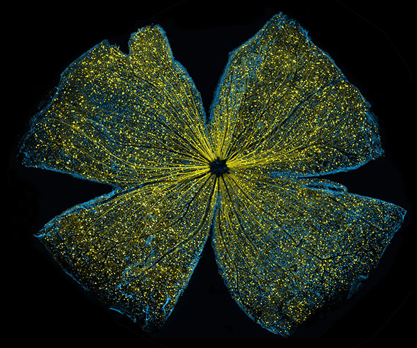
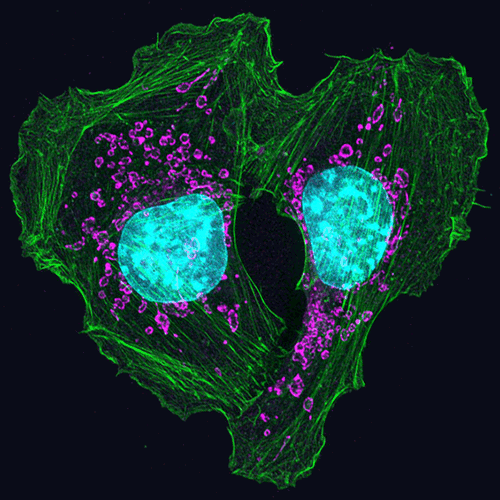
This heart-shaped image shows two mouse skin cancer cells connected to each other with actin, a protein that is part of the cellular skeleton. Researchers use mouse cells like these to tease out the molecular methods that cancer uses to invade new tissues in the body. It turns out that actin plays an essential role.

Cells can move as a collective, or independently. Movement of an individual cell requires a series of carefully controlled steps. Among them, a cell must break contacts with its neighbor cells and change its connections to the proteins and fibers around it. In addition, it must sense and follow a chemical path through the tissue it lies in. To do this, a cell changes shape, molding its membrane into flaps or feet called protrusions reaching in the direction it is traveling. Actin, among a variety of other molecules, is involved in all of these steps, but especially the shape change, when it gathers inside the cell membrane to help form the protrusions. Continue reading “Actin’s Many Roles”

Cool Image: Inside a Biofilm Build-up

1 comment
A growing Vibrio cholerae biofilm.

A growing Vibrio cholerae biofilm. Each slightly curved comma shape represents an individual bacterium from assembled confocal microscopy images. Different colors show each bacterium’s position in the biofilm in relation to the surface on which the film is growing. Credit: Jing Yan, Ph.D., and Bonnie Bassler, Ph.D., Department of Molecular Biology, Princeton University, Princeton, NJ.

Bacteria use many methods to overcome threats in their environment. One of these ways is forming colonies called biofilms on surfaces of objects. Often appearing like the bubble-shaped fortress represented in this image, biofilms enable bacteria to withstand attacks, compete for space and survive fluctuations in nutrient supply. Bacteria aggregated within biofilms inside our bodies, for example, resist antibiotic therapy more effectively than free swimming cells, making infections difficult to treat. On the other hand, biofilms are also useful for making microbial fuel cells and for waste-water treatment. Learning how biofilms work, therefore, could provide essential tools in our ongoing battle against disease-causing agents and in our efforts to harness beneficial bacterial behaviors. Researchers are now using new imaging techniques to watch how biofilms grow, cell by cell, and to identify more effective ways of disrupting or fostering them.

Until now, poor imaging resolution meant that scientists could not see what individual bacteria in the films are up to as the biofilms grow. The issue is that bacteria are tiny, making imaging each cell, as well as the ability to distinguish each cell in the biofilm community, problematic. Continue reading “Cool Image: Inside a Biofilm Build-up”

Cool Image: Adding Color to the Gray World of Electron Microscopy

1 comment

Color electron micrograph of an endosome, a cell organelle. Credit: Ranjan Ramachandra, UCSD

As his Christmas gift to himself each year, the late biochemist Roger Tsien treated himself to two weeks of uninterrupted research in his lab. This image is a product of those annual sojourns. While it may look like a pine wreath dotted with crimson berries, it is in fact one of the world’s first color electron micrographs—and the method used to create it may dramatically advance cell imaging.

Electron microscopy (EM) is a time-honored technique for visualizing cell structures that uses beams of accelerated electrons to magnify objects up to 10 million times their actual size. Standard EM images are in grayscale and any color is added in with computer graphics programs after the image is made. With their new technique, Tsien, who received a Nobel Prize for his development of green fluorescent protein into a tool for visualizing details in living cells using light microscopes, and his colleagues have found a way to incorporate color labeling directly into EM. Continue reading “Cool Image: Adding Color to the Gray World of Electron Microscopy”

Metals in Medicine

4 comments

An exhibit called “Minerals in Medicine” opened at the NIH Clinical Center last month (see slideshow). The display features a fascinating overview of how dozens of minerals are used to create drugs and medical instruments useful in treating disease and maintaining health. The minerals ranged from commonplace ones like quartz, which is used to make medical instruments, to more exotic ones like hubnerite, a source of the metal tungsten, which is used in radiation shielding.

Inspired by this collection, which is co-sponsored by NIH and the Smithsonian Institution, we highlight here examples of “Metals in Medicine.”

Copper and Fat Metabolism

Fluorescent imaging of copper in white fat cells from mice.

Fluorescent imaging of copper in white fat cells from mice. The left panel shows fat cells with normal levels of copper, and the right panel shows fat cells deficient in copper. Credit: Lakshmi Krishnamoorthy and Joseph Cotruvo Jr., University of California, Berkeley.

What does a metal like copper have to do with our ability to breakdown fat? Researchers explored this question by observing mice with Wilson’s disease—a rare, inherited condition that causes copper to accumulate in the liver, brain and other vital organs. The mice with the condition usually have larger deposits of fat compared to healthy mice. To confirm that fat metabolism is somehow compromised in these mice, the researchers treated them with a drug that induces the breakdown of fat. And indeed they found that less fat was metabolized in mice with the disease.

In an effort to investigate what role copper may be playing in fat metabolism, the researchers examined adipose tissue, or fat, cells under a microscope to track the metal’s interactions with various proteins in the cell. They discovered that copper inhibits an enzyme called PDE3. This enzyme usually prevents another enzyme called cAMP from helping to break down fat. The researchers concluded that copper actually promotes fat metabolism. This work shows that transition metal nutrients can play signaling roles, which has been previously thought to be restricted to alkali and alkaline earth metals like sodium, potassium and calcium.

Continue reading “Metals in Medicine”

NIGMS Is on Instagram!

0 comments

Science is beautiful.

For several years, we’ve used this blog to highlight pictures we think are cool, scientifically relevant and visually striking. The images were created by NIGMS-funded researchers in the process of doing their research. Many come from our Life: Magnified collection, which features dozens of stunning photos of life, close-up. We’ll continue to bring you interesting images and information here on Biomedical Beat, but if you can’t get enough of them, we have a new way to share our visual content with you: Instagram.

We’re pleased to announce the launch of our NIGMS Instagram account. We’ll highlight gorgeous images, and bring you the science behind them—straight from our mobile device to yours. Instagram lets us label our images with subject-specific hashtags. You can find our pictures by going on Instagram and searching for #NIGMS. If you’re already an Instagram user, you can follow us @NIGMS_NIH. You can even see our page using a web browser at https://www.instagram.com/nigms_nih/ Exit icon. Let us know what you think!

Also, if you have any stunning images or videos that relate to scientific areas supported by NIGMS, please send them to us. They might end up on our Instagram feed!

See those finger-like projections? They are called villi. This image shows the small intestine, where most of the nutrients from the food we eat are absorbed into the bloodstream. The villi increase the organ’s surface area, making nutrient absorption more efficient. Credit: National Center for Microscopy and Imaging Research.
#science Exit icon #biology Exit icon #research Exit icon #cells Exit icon #cellbiology Exit icon #microscopy Exit icon #nigms Exit icon #scienceisbeautiful Exit icon #sciart Exit icon #nigms Exit icon #nih Exit icon #NCMIR Exit icon #UCSD Exit icon #smallbowel Exit icon  #nutrients Exit icon #nutrient Exit icon #anatomyphysiology Exit icon

We need zinc. It’s an essential nutrient for growth and development, fending off invading microbes, healing injuries, and all sorts of cellular processes. We get the mineral through our diet, but people in certain parts of the world don’t get enough. Researchers study how plants acquire and process zinc, hoping to find ways to increase the nutrient in food crops. Using synchrotron X-ray fluorescence technology, scientists created this heat map of zinc in a leaf from a plant called Arabidopsis thaliana (zinc levels from lowest to highest: blue, green, red, white). Credit: Suzana Car and Mary Lou Guerinot, Dartmouth College.
#science Exit icon #biology Exit icon #research Exit icon #botany Exit icon #plant Exit icon #arabidopsis Exit icon #zinc Exit icon #microscopy Exit icon #synchrotron Exit icon #x-ray Exit icon #modelorganism Exit icon #leaf Exit icon #heatmap Exit icon #scienceisbeautiful Exit icon #nigms Exit icon #nih Exit icon

Beautiful brain! This image shows the cerebellum, which is the brain’s locomotion control center. Every time you shoot a basketball, tie your shoe or chop an onion, your cerebellum fires into action. Found at the base of your brain, the cerebellum is a single layer of tissue with deep folds like an accordion. People with damage to this region of the brain often have difficulty with balance, coordination and fine motor skills. Credit: Tom Deerinck and Mark Ellisman, NCMIR
#science Exit icon #biology Exit icon #research Exit icon #cerebellum Exit icon #neuroscience Exit icon #neurobiology Exit icon #brain Exit icon #brains Exit icon #brainimages Exit icon #neuroanatomy Exit icon #microscopy Exit icon #scienceisbeautiful Exit icon #sciart Exit icon #nigms Exit icon #nih Exit icon #NCMIR Exit icon #UCSD Exit icon

Scientists can learn a lot by studying pigment cells, which give animals their colorful skins, eyes, hair and scales. We can even gain insight into skin cancers, like melanoma, that originate from pigment cells. Pigment cells can form all sorts of patterns, like these stripes on the fin of a pearl danio, a type of tropical minnow. Credit: David Parichy, University of Washington.
#biology Exit icon #marinebiology Exit icon #marinescience Exit icon #cell Exit icon #pigmentcell Exit icon #xanthophore Exit icon #science Exit icon #research Exit icon #cellbiology Exit icon #microscopy Exit icon #scienceisbeautiful Exit icon #scienceiscool Exit icon #zebrafish Exit icon #universityofwashington Exit icon

Lighting Up the Promise of Gene Therapy for Glaucoma

0 comments
Retinal ganglion cells in the mouse.

Retinal ganglion cells in the mouse retina that do (bright, yellow spots throughout) and do not (blue streaks, mostly along the edges) contain a specific gene that scientists introduced with a virus. Credit: Kenyoung (“Christine”) Kim, Wonkyu Ju and Mark Ellisman, National Center for Microscopy and Imaging Research, University of California, San Diego.

What looks like the gossamer wings of a butterfly is actually the retina of a mouse, delicately snipped to lay flat and sparkling with fluorescent molecules. Researchers captured this image while investigating the promise of gene therapy for glaucoma, a progressive eye disease. It all happened at the National Center for Microscopy and Imaging Research Exit icon (NCMIR) at the University of California, San Diego.

Glaucoma is the leading cause of irreversible blindness. It is characterized by the slow, steady death of certain nerve cells in the retina. If scientists can prevent the death of these cells, which are called retinal ganglion cells, it might be possible to slow the progression of glaucoma. Some researchers are examining the possibility of using gene therapy to do just that.

A major challenge of gene therapy is finding a way to get therapeutic genes into the right cells without damaging the cells in any way. Scientists have had success using a non-disease-causing virus (adeno-associated serotype 2) for this task. Continue reading “Lighting Up the Promise of Gene Therapy for Glaucoma”

Exploring the Evolution of Spider Venom to Improve Human Health

1 comment
Brown recluse

Female brown recluse spider. Credit Matt Bertone, North Carolina State University.

This Halloween, you’re not likely to see many trick-or-treaters dressed as spiders. Google Trends pegs “Spider” as the 87th most searched-for Halloween costume, right between “Hippie” and “The Renaissance.” But don’t let your guard down. Spiders are everywhere.

“I grew up on a farm in Indiana and had the luxury of exploring and turning over rocks and being curious. Any feelings of being grossed out by spiders were rapidly replaced by my feelings of awe for how amazing and diverse these creatures are.”– Greta Binford”

More than 46,000 species of spiders creepy crawl across the globe, on every continent except Antarctica. Each species produces a venom composed of an average of 500 distinct toxins, putting the conservative estimate of unique venom compounds at more than 22 million. This staggering diversity of venoms, collectively referred to as the venome, has only begun to be explored. Continue reading “Exploring the Evolution of Spider Venom to Improve Human Health”

Newly Identified Cell Wall Construction Workers: A Novel Antibiotic Target?

0 comments
SEDS

A family of proteins abbreviated SEDS (bright, pink) help build bacterial cell walls, so they are a potential target for new antibiotic drugs. Credit: Rudner lab, Harvard Medical School.

Scientists have identified a new family of proteins that, like the targets of penicillin, help bacteria build their cell walls. The finding might reveal a new strategy for treating a range of bacterial diseases.

The protein family is nicknamed SEDS, because its members help control the shape, elongation, division and spore formation of bacterial cells. Now researchers have proof that SEDS proteins also play a role in constructing cell walls. This image shows the movement of a molecular machine that contains a SEDS protein as it constructs hoops of bacterial cell wall material.

Any molecule involved in building or maintaining cell walls is of immediate interest as a possible target for antibiotic drugs. That’s because animals, including humans, don’t have cell walls—we have cell membranes instead. So disabling cell walls, which bacteria need to survive, is a good way to kill bacteria without harming patients.

This strategy has worked for the first antibiotic drug, penicillin (and its many derivatives), for some 75 years. Now, many strains of bacteria have evolved to resist penicillins—and other antibiotics—making the drugs less effective.

According to the Centers for Disease Control and Prevention, drug-resistant strains of bacteria Exit icon infect at least 2 million people, killing more than 20,000 of them in the U.S. every year. Identifying potential new drug targets, like SEDS proteins, is part of a multi-faceted approach to combating drug-resistant bacteria.

Pigment Cells: Not Just Pretty Colors

2 comments

If you’ve ever visited an aquarium or snorkeled along a coral reef, you’ve witnessed the dazzling colors and patterns on tropical fish. The iridescent stripes and dots come from pigment cells, which also tint skin, hair and eyes in all kinds of animals, including humans. Typically, bright colors help attract mates, while dull ones provide camouflage. In humans, pigment helps protect skin from DNA-damaging UV light.

Researchers study cellular hues not only to decipher how they color our world, but also to understand skin cancers that originate from pigment cells. Some of these researchers work their way back, developmentally speaking, to focus on the type of cell, known as a neural crest cell, that is the precursor of pigment cells.

Present at the earliest stages of development, neural crest cells migrate throughout an embryo and transform into many different types of cells and tissues, including nerve cells, cartilage, bone and skin. The images here, from research on neural crest cells in fish and salamanders, showcase the beauty and versatility of pigment cells in nature’s palette.

Xanthophores
Pigment cells called xanthophores, shown here in the skin of the popular laboratory animal zebrafish, glow brightly under light. Credit: David Parichy, University of Washington.
Melanocytes
Dark pigment cells, called melanocytes, like these in pearl danio, a tropical minnow and relative of zebrafish, assemble in skin patterns that allow the animals to blend into their surroundings or attract mates. Credit: David Parichy, University of Washington.
Fin of pearl danio
Pigment cells can form all sorts of patterns, like these stripes on the fin of pearl danio. Credit: David Parichy, University of Washington.
Salamander skin
Pigment cells arise from neural crest cells. Here, pigment cells can be seen migrating in the skin of a salamander where they will form distinct color patterns. Credit: David Parichy, University of Washington.


A Labor Day-Themed Collection: Hard-Working Cell Structures

2 comments

Hard labor might be the very thing we try to avoid on Labor Day. But our cells and their components don’t have the luxury of taking a day off. Their non-stop work is what keeps us going and healthy.

Scientists often compare cells with small factories. Just like a factory, a cell contains specialized compartments and machines—including organelles and other structures—that each play their own roles in getting the job done. In the vignettes below, we give a shout out to some of these tireless cellular workers.

Energy Generators
Credit: Thomas Deerinck, National Center for Microscopy and Imaging Research
Mitochondria are the cell’s power plants. They convert energy from food into a molecule called ATP that fuels virtually every process in the cell. As shown here, mitochondria (brown) often have distinct, oblong shapes. Like most other organelles, mitochondria are encased in an outer membrane. But they also have an inner membrane that folds many times, increasing the area available for energy production. In addition, mitochondria store calcium ions, help make hemoglobin—the vital iron-containing protein that allows red blood cells to carry oxygen—and even take part in producing some hormones. Defects in mitochondria can lead to a host of rare but often incurable diseases that range from mild to devastating. Researchers are studying mitochondria to better understand their manifold jobs in the cell and to find treatments for mitochondrial diseases.

Continue reading “A Labor Day-Themed Collection: Hard-Working Cell Structures”