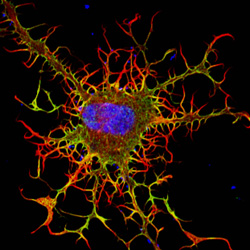
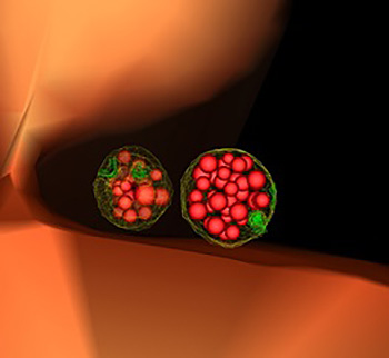
In large offices, mailroom workers read the labels on incoming letters and packages to sort and deliver them and dispose of junk mail. In cells, these tasks—as well as importing food and other materials—fall to small cellular sacs called endosomes. Acting as mailroom staff, endosomes sort and deliver nutrients and building blocks, like amino acids, fat and sugars, to their proper destinations, and send cellular junk, like damaged proteins, to trash processors, such as vacuoles or lysosomes. Continue reading “The Cell’s Mailroom”
Tag: Cool Images
A World Without Pain
You glide across an icy canyon where you meet smiling snowmen, waddling penguins and a glistening river that winds forever. You toss snowballs, hear them smash against igloos, then watch them explode in vibrant colors.
Back in the real world, a dentist digs around your mouth to remove an impacted tooth, a procedure that really, really hurts. Could experiencing a “virtual” world distract you from the pain? NIGMS grantees David Patterson and Hunter Hoffman show it can.
Patterson, a psychologist at the University of Washington (UW) in Seattle, and Hoffman, a UW cognitive psychologist, helped create the virtual reality program “Snow World” in an effort to reduce excessive pain experienced by burn patients. However, the researchers expect Snow World to help alleviate all kinds of pain, including pain experienced during dental procedures. Continue reading “A World Without Pain”
Cool Images: An Independence Day-Inspired Collection
@media screen and (max-width: 480px) {
.halloween-out {
width: 96.5%;
}
.halloween-out .halloween-in img {
padding-top: 42px;
padding-right: 12px;
}
}
@media screen and (max-width: 320px) {
.halloween-out {
width: 96%;
}
}
In case you missed the fireworks this weekend, we’ve put together a collection of firework-like images from basic research studies.
Viral Views: New Insights on Infection Strategies
The following images show a few ways in which cutting-edge research tools are giving us clearer views of viruses—and possible ways to disarm them. The examples, which highlight work involving HIV and the coronavirus, were funded in part by our Biomedical Technology Research Resources program.
Uncloaking HIV’s Camouflage
To sneak past our immune defenses and infect human cells, HIV uses a time-honored strategy—disguise. The virus’ genome is enclosed in a protein shell called a capsid (on left) that’s easily recognized and destroyed by the human immune system. To evade this fate, the chrysalis-shaped capsid cloaks itself with a human protein known as cyclophilin A (in red, on right). Camouflaged as human, the virus gains safe passage into and through a human cell to deposit its genetic material in the nucleus and start taking control of cellular machinery.
Biomedical and technical experts teamed up to generate these HIV models at near-atomic resolution. First, structural biologists at the Pittsburgh Center for HIV Protein Interactions used a technique called cryo-electron microscopy (cryo-EM) to get information on the shape of an HIV capsid as well as the capsid-forming proteins’ connections to each other and to cyclophilin A. Then experts at the Resource for Macromolecular Modeling and Bioinformatics fed the cryo-EM data into their visualization and simulation programs to computationally model the physical interactions among every single atom of the capsid and the cyclophilin A protein. The work revealed a previously unknown site where cyclophilin A binds to the capsid, offering new insights on the biology of HIV infection. Continue reading “Viral Views: New Insights on Infection Strategies”
Visualizing Skin Regeneration in Real Time
Zebrafish, blue-and-white-striped fish that are about 1.5 inches long, can regrow injured or lost fins. This feature makes the small fish a useful model organism for scientists who study tissue regeneration.
To better understand how zebrafish skin recovers after a scrape or amputation, researchers led by Kenneth Poss of Duke University tracked thousands of skin cells in real time. They found that lifespans of individual skin cells on the surface were 8 to 9 days on average and that the entire skin surface turned over in 20 days.
The scientists used an imaging technique they developed called “Skinbow,” which essentially shows the fish’s outer layer of skin cells in a spectrum of colors when viewed under a microscope. Skinbow is based on a technique created to study nerve cells in mice, another model organism.
The research team’s color-coded experiments revealed several unexpected cellular responses during tissue repair and replacement. The scientists plan to incorporate additional imaging techniques to generate an even more detailed picture of the tissue regeneration process.
The NIH director showcased the Skinbow technique and these images on his blog, writing: “You can see more than 70 detectable Skinbow colors that make individual cells as visually distinct from one another as jellybeans in a jar.”
This work was funded in part by NIH under grant R01GM074057.
Cool Image: A Circadian Circuit
Feeling sleepy and dazed after the switch to daylight savings time this weekend? Your internal clocks are probably a little off and need some time to adjust.
Researchers have been studying biological clocks for decades to figure out how they control circadian rhythms, the natural 24-hour pattern of physical, mental and behavioral changes that affect sleep, appetite and metabolism. Knowing more about what makes our clocks tick could help researchers develop better therapies for sleep problems, metabolic conditions and other disorders associated with mistimed internal clocks. Continue reading “Cool Image: A Circadian Circuit”
A Heart-Shaped Protein
From cookies and candies to balloons and cards, heart-shaped items abound this time of year. They’re even in our blood. It turns out that the most abundant protein molecule in blood plasma—serum albumin (SA)—is shaped very much like a heart. Continue reading “A Heart-Shaped Protein”
Cool Images: A Holiday-Themed Collection
Here are some images from our gallery that remind us of the winter holidays—and showcase important findings and innovations in biomedical research.
Ribbons and Wreaths
This wreath represents the molecular structure of a protein, Cas4, which is part of a system, known as CRISPR, that bacteria use to protect themselves against viral invaders. The green ribbons show the protein’s structure, and the red balls show the location of iron and sulfur molecules important for the protein’s function. Scientists have harnessed Cas9, a different protein in the bacterial CRISPR system, to create a gene-editing tool known as CRISPR-Cas9. Using this tool, researchers can study a range of cellular processes and human diseases more easily, cheaply and precisely. Last week, Science magazine recognized the CRISPR-Cas9 gene-editing tool as the “breakthrough of the year.”
Cool Image: Tracing Proteins in Action
Looking like necklaces stacked on a dresser, these bright, amorphous loops show the outlines of yeast proteins that make up the spindle pole, a cellular component found in organisms as diverse as yeast and humans. Each cell starts with a single spindle pole, which must somehow duplicate to form the pair that works together to pull matching chromosomes apart during cell division. Scientists don’t completely understand how this duplication occurs, but they do know that errors in spindle pole copying can lead to a number of health conditions, including cancer.
Continue reading “Cool Image: Tracing Proteins in Action”Cool Images: A Halloween-Inspired Cell Collection
As Halloween approaches, we turned up some spectral images from our gallery. The collection below highlights some spooky-sounding—but really important—biological topics that researchers are actively investigating to spur advances in medicine.
Continue reading “Cool Images: A Halloween-Inspired Cell Collection”