Author: Kathryn Calkins

Headshot of Kathryn Calkins.

Posts by Kathryn Calkins

The Irresistible Resistome: How Infant Diapers Might Help Combat Antibiotic Resistance (sort of)

5 comments
Gautam Dantas
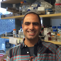
Credit: Pablo Tsukayama, Ph.D.,
Washington University School of Medicine
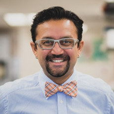
Gautam Dantas
Born: Mumbai, India
Most proud of: His family, which brings him joy and pride
Favorite lab tradition: OOFF! Official Optional Formal Fridays, when members of his lab can dress up, eat bread—made in the lab’s own bread machine—and drink beer and wine together at the end of the day
When not in the lab, he: Enjoys home brewing, pickling and canning, and spending time with his wife and children. He also attends musical performances, including those of his wife, who sings in the St. Louis Symphony Chorus
Advice to aspiring scientists: Pursue hobbies, take risks, explore beyond your comfort zone. “You can do a Ph.D., but also have other experiences.” He says his own outside activities refine his focus in the lab, keep him grounded and help him be an empathetic mentor to his students. Plus, he met his wife while singing in the chorus of Macalester College in St. Paul, Minnesota

When I Grow Up…

Gautam Dantas remembers the day in 10th grade when he first wanted to be a scientist. It was the day he had a new biology teacher, a visiting researcher from the U.S. The teacher passionately described his own biochemical studies of how organisms live together in communities. By the end of the class, Dantas had resolved to earn a Ph.D. in biochemistry.

He ended up doing much more—gaining expertise in computational biology, protein design and synthetic biology. He now combines his skills and knowledge in multifaceted research that spans four departments at Washington University in St. Louis. His goal: to better understand and help combat a vital public health threat—drug-resistant bacteria.

“Our motivation is that we are living in the antibiotic era, and antibiotic resistance is getting out of control,” Dantas says. “We have very few new antibiotics we can use, so we’re kind of scrambling [to find new ways to treat bacterial diseases].”

His research focuses on one of the groups most vulnerable to bacterial infections—newborn babies.

According to his lab’s website Exit icon, the research is “at the interface of microbial genomics, ecology, synthetic biology, and systems biology,” and it aims “to understand, harness, and engineer the biochemical processing potential of microbial communities.” They do it by scrounging around in infant diapers.

Antibiotic Angst

Since their introduction in the 1940s, antibiotic drugs have saved countless lives. Simultaneously, they weeded out strains of bacteria easily killed by the drugs, allowing drug-resistant strains to thrive. Every year, at least 2 million people in the U.S. become infected and at least 23,000 die from drug-resistant bacteria, according to the Center for Disease Control and Prevention. Continue reading “The Irresistible Resistome: How Infant Diapers Might Help Combat Antibiotic Resistance (sort of)”

The ECM: A Dynamic System for Moving Our Cells

2 comments

In part I of this series, we mentioned that the extracellular matrix (ECM) makes our tissues stiff or squishy, solid or see-through. Here, we reveal how the ECM helps body cells move around, a process vital for wounds to heal and a fetus to grow.

Sealing and Healing Wounds

MMPs are essential for closing wounds. Credit: Stock image.

When we get injured, the first thing our body does is to form a blood clot to stop the bleeding. Skin cells then start migrating into the wound to close the cut. The ECM is essential for this step, creating a physical support structure—like a road or train track—over which skin cells travel to seal the injured spot.

The ECM is made up of a host of proteins produced before and after injury. Some other proteins called matrix metalloproteinases (MMPs) also crowd into wounds. Because humans have so many different MMPs—a full 24 of them!—it’s been difficult for scientists to figure out what roles, if any, the proteins play in healing scrapes and cuts.

Continue reading “The ECM: A Dynamic System for Moving Our Cells”

A Labor Day-Themed Collection: Hard-Working Cell Structures

2 comments

Hard labor might be the very thing we try to avoid on Labor Day. But our cells and their components don’t have the luxury of taking a day off. Their non-stop work is what keeps us going and healthy.

Scientists often compare cells with small factories. Just like a factory, a cell contains specialized compartments and machines—including organelles and other structures—that each play their own roles in getting the job done. In the vignettes below, we give a shout out to some of these tireless cellular workers.

Energy Generators
Credit: Thomas Deerinck, National Center for Microscopy and Imaging Research
Mitochondria are the cell’s power plants. They convert energy from food into a molecule called ATP that fuels virtually every process in the cell. As shown here, mitochondria (brown) often have distinct, oblong shapes. Like most other organelles, mitochondria are encased in an outer membrane. But they also have an inner membrane that folds many times, increasing the area available for energy production. In addition, mitochondria store calcium ions, help make hemoglobin—the vital iron-containing protein that allows red blood cells to carry oxygen—and even take part in producing some hormones. Defects in mitochondria can lead to a host of rare but often incurable diseases that range from mild to devastating. Researchers are studying mitochondria to better understand their manifold jobs in the cell and to find treatments for mitochondrial diseases.

Continue reading “A Labor Day-Themed Collection: Hard-Working Cell Structures”

CRISPR Serves Up More than DNA

0 comments
Marine bacterium Marinomonas mediterranea
The marine bacterium Marinomonas mediterranea uses a CRISPR system to spot invading RNAs and store a memory of the invasion event in its genome. Research team member Antonio Sanchez-Amat was the first to isolate and characterize this bacterial species. Credit: Antonio Sanchez-Amat, University of Murcia.

A new study has added another twist to the CRISPR story. As we’ve highlighted in several recent posts, CRISPR is an immune system in bacteria that recognizes and destroys viral DNA and other invading DNA elements, such as transposons. Scientists have adapted CRISPR into an indispensable gene-editing tool now widely used in both basic and applied research.

Many previously described CRISPR systems detect and cut viral DNA, insert the DNA pieces into the bacterial genome and then use them as molecular “mug shots” to flag and destroy the virus if it attacks again. But various viruses use RNA, not DNA, as genetic material. Although research has shown that some CRISPR systems also can target RNA, how these systems can archive harmful RNA encounters in the bacterial genome was unknown. Continue reading “CRISPR Serves Up More than DNA”

Finding Adventure: Blake Wiedenheft’s Path to Gene Editing

1 comment
Blake Wiedenheft
Blake Wiedenheft
Grew up in: Fort Peck, Montana
Fields: Microbiology, biochemistry, structural biology
Job site: Montana State University
Secret talent: Being a generalist; enjoying many different subjects and activities
When not in the lab, he’s: Running, biking, skiing or playing scrabble with his grandmother

Scientific discoveries are often stories of adventure. This is the realization that set Blake Wiedenheft on a path toward one of the hottest areas in biology.

His story begins in Montana, where he grew up and now lives. Always exploring different interests, Wiedenheft decided in his final semester at Montana State University (MSU) in Bozeman to volunteer for Mark Young, a scientist who studies plant viruses. Even though he majored in biology, Wiedenheft had spent little time in a lab and hadn’t even considered research as a career option. Continue reading “Finding Adventure: Blake Wiedenheft’s Path to Gene Editing”

A Heart-Shaped Protein

1 comment
Structure of the serum albumin protein

The structure of the serum albumin protein is shaped like a heart. Credit: Wladek Minor, University of Virginia.

From cookies and candies to balloons and cards, heart-shaped items abound this time of year. They’re even in our blood. It turns out that the most abundant protein molecule in blood plasma—serum albumin (SA)—is shaped very much like a heart. Continue reading “A Heart-Shaped Protein”

Cool Images: A Holiday-Themed Collection

1 comment

Here are some images from our gallery that remind us of the winter holidays—and showcase important findings and innovations in biomedical research.

Ribbons and Wreaths

Wreath

This wreath represents the molecular structure of a protein, Cas4, which is part of a system, known as CRISPR, that bacteria use to protect themselves against viral invaders. The green ribbons show the protein’s structure, and the red balls show the location of iron and sulfur molecules important for the protein’s function. Scientists have harnessed Cas9, a different protein in the bacterial CRISPR system, to create a gene-editing tool known as CRISPR-Cas9. Using this tool, researchers can study a range of cellular processes and human diseases more easily, cheaply and precisely. Last week, Science magazine recognized the CRISPR-Cas9 gene-editing tool as the “breakthrough of the year.”

Continue reading “Cool Images: A Holiday-Themed Collection”

Bacterial Biofilms: A Charged Environment

1 comment
Bacillus subtilis biofilm
A Bacillus subtilis biofilm grown in a Petri dish. Credit: Süel Lab, UCSD.

Last summer, we shared findings from Gürol Süel Exit icon and colleagues at the University of California, San Diego, that bacterial cells in tight-knit microbial communities called biofilms expand in a stop-and-go pattern. The researchers concluded that this pattern helps make food at the nutrient-rich margin available to the cells in the starved center, but they didn’t know how. They’ve now shown that the cells use electrochemical signaling to communicate and cooperate with each other.

Because nutrients and other signals cells use to sense each other and their environment move rather slowly, the researchers looked for a faster, more active communication system in biofilms of the bacterium B. subtilis. They focused on electrical signaling via potassium, a positively charged ion that, for example, our nerve and muscle cells use to send or receive signals. Continue reading “Bacterial Biofilms: A Charged Environment”

Cool Images: A Halloween-Inspired Cell Collection

3 comments

As Halloween approaches, we turned up some spectral images from our gallery. The collection below highlights some spooky-sounding—but really important—biological topics that researchers are actively investigating to spur advances in medicine.

Cell Skeleton
Fibroblast
The cell skeleton, or cytoskeleton, is the framework that gives a cell its shape, helps it move and keeps its contents organized for proper function. A cell that lacks a cytoskeleton becomes misshapen and immobile. This fibroblast, a cell that normally makes connective tissues and travels to the site of a wound to help it heal, is lacking a cytoskeleton. Researchers have associated faulty cytoskeletons and resulting abnormal cell movement with birth defects and weakened immune system functioning. See fibroblasts with healthy skeletons.

Continue reading “Cool Images: A Halloween-Inspired Cell Collection”

Cool Image: DNA Origami

0 comments
Computer-generated sketch of a DNA origami folded into a flower-and-bird structure.
A computer-generated sketch of a DNA origami folded into a flower-and-bird structure. Credit: Hao Yan, Arizona State University.

This image of flowers visited by a bird is made of DNA, the molecule that provides the genetic instructions for making living organisms. It shows the latest capability of a technique called DNA origami to precisely twist and fold DNA into complex arrangements, which might find future use in biomedical applications.

Continue reading “Cool Image: DNA Origami”