Tag: Cellular Imaging

Zinc’s Role in Healthy Fertilization

0 comments
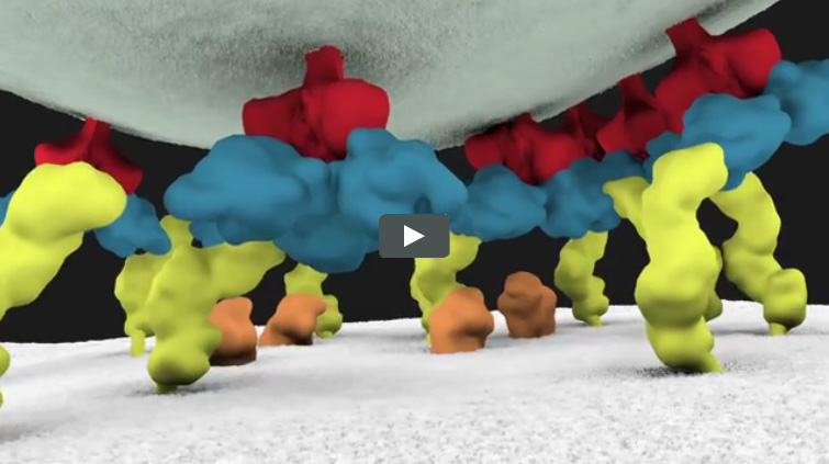
Screen shot of the video
Fluorescent sensors at the cell surface show zinc-rich packages being released from the egg during fertilization. Credit: Northwestern Visualization.

Whether aiding in early growth and development, ensuring a healthy nervous system or guarding the body from illness, zinc plays an important role in the human body.

Husband-and-wife team, Thomas O’Halloran Exit icon and Teresa Woodruff, plus other researchers at Northwestern University, evaluated the role that zinc plays in healthy fertilization Exit icon. The study revealed how mouse eggs gather and release billions of zinc atoms at once in events called zinc sparks. These fluxes in zinc concentration are essential in regulating the biochemical processes that facilitate the egg-to-embryo transition.

The scientists developed a series of techniques to determine the amount and location of zinc atoms during an egg cell’s maturation and fertilization as well as in the following two hours. Special imaging methods allowed the researchers to also visualize the movement of zinc sparks in three dimensions. Continue reading “Zinc’s Role in Healthy Fertilization”

A Bright New Method for Rapidly Screening Cancer Drugs

0 comments
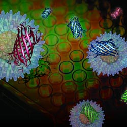
Illustration of red, green and blue fluorescent proteins.
Chemists have devised a new approach to screening cancer drugs that uses gold nanoparticles with red, green and blue outputs provided by fluorescent proteins. Credit: University of Massachusetts Amherst.

Scientists may screen billions of chemical compounds before uncovering the few that effectively treat a disease. But identifying compounds that work is just the first step toward developing a new therapy. Scientists then have to determine exactly how those compounds function.

Different cancer therapies attack cancer cells in distinct ways. For example, some drugs kill cancer cells by causing their outer membranes to rapidly rupture in a process known as necrosis. Others cause more subtle changes to cell membranes, which result in a type of programmed cell death known as apoptosis.

If researchers could distinguish the membrane alterations of chemically treated cancer cells, they could quickly determine how that chemical compound brings about the cells’ death. A new sensor developed by a research team led by Vincent Rotello of the University of Massachusetts Amherst can make these distinctions in minutes. Continue reading “A Bright New Method for Rapidly Screening Cancer Drugs”

Illuminating Biology

0 comments

This time of year, lights brighten our homes and add sparkle to our holidays. Year-round, scientists funded by the National Institutes of Health use light to illuminate important biological processes, from the inner workings of cells to the complex activity of the brain. Here’s a look at just a few of the ways new light-based tools have deepened our understanding of living systems and set the stage for future medical advances.

RSV infected cell
A new fluorescent probe shows viral RNA (red) in an RSV-infected cell. Credit: Eric Alonas and Philip Santangelo, Georgia Institute of Technology and Emory University.

Visualizing Viral Activity

What looks like a colorful pattern produced as light enters a kaleidoscope is an image of a cell infected with respiratory syncytial virus (RSV) lit up by a new fluorescent probe called MTRIPS (multiply labeled tetravalent RNA imaging probes).

Although relatively harmless in most children, RSV can lead to bronchitis and pneumonia in others. Philip Santangelo of the Georgia Institute of Technology and Emory University, along with colleagues nationwide, used MTRIPS to gain a closer look at the life cycle of this virus.

Once introduced into RSV-infected cells, MTRIPS latched onto the genetic material of individual viral particles (in the image, red), making them glow. This enabled the researchers to follow the entry, assembly and replication of RSV inside the living cells. Continue reading “Illuminating Biology”

Field Focus: Bringing Biology Into Sharper View with New Microscopy Techniques

0 comments
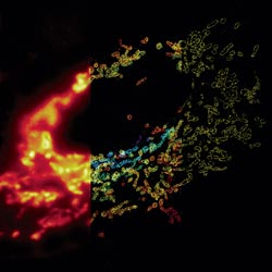
Composite image of mitochondria in a cell
In this composite image of mitochondria in a cell, the left panel shows a conventional optical microscopy image, the middle panel shows a three-dimensional (3-D) STORM image with color indicating depth, and the right panel shows a cross-section of the 3-D STORM image. Credit: Xiaowei Zhuang laboratory, Howard Hughes Medical Institute, Harvard University. View larger image.

Much as a photographer brings distant objects into focus with a telephoto lens, scientists can now see previously indistinct cellular components as small as a few billionths of a meter (nanometers). By overcoming some of the limitations of conventional optical microscopy, a set of techniques known as super-resolution fluorescence microscopy has changed once-blurry images of the nanoworld into well-resolved portraits of cellular architecture, with details never seen before in biology. Reflecting its importance, super-resolution microscopy was recognized with the 2014 Nobel Prize in chemistry.

Using the new techniques, scientists can observe processes in living cells across space and time and study the movements, interactions and roles of individual molecules. For instance, they can identify and track the proteins that allow a virus to invade a cell or those that enable tumor cells to migrate to distant parts of the body in metastatic cancer. The ability to analyze individual molecules, rather than collections of molecules, allows scientists to answer longstanding questions about cellular mechanisms and behavior, such as how cells move along a surface or how certain proteins interact with DNA to regulate gene activity. Continue reading “Field Focus: Bringing Biology Into Sharper View with New Microscopy Techniques”

Stem Cells Do Geometry

2 comments
Human embryonic cells
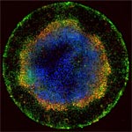
As seen under a microscope, human embryonic cells (colored dots) confined to circles measuring 1 millimeter across start to specialize and form distinct layers similar to those seen in early development. Credit: Aryeh Warmflash, Rockefeller University. View larger image

Each fluorescent point of light making up the multicolored rings in this image is an individual human embryonic cell in the early stages of development. Scientists seeking to understand the molecular cues responsible for early embryonic patterning found that human embryonic cells confined to areas of precisely controlled size and shape begin to specialize, migrate and organize into distinct layers just as they would under natural conditions.

Read the Inside Life Science article to learn more about this research, which has opened a new window for studying early development and could advance efforts aimed at using human stem cells to replace diseased cells and regenerate lost or injured body parts.

Steering Cells Down the Right Path

1 comment
In these time-lapse videos (no longer available) of 60 images taken over an hour, cell receptors move around the cell surface in search of the missing signal that will tell the cell where to go (top video). Once the receptors locate the signal (bottom video), they stay put in the region of the cell membrane that is closest to the signal. Credit: David Sherwood, Duke University.

Even traveling cells need help with directions. In fact, it’s crucial. For processes such as wound healing and organ development to take place, cells must be able to efficiently move throughout organisms. Receptor proteins on a cell’s surface rely on navigational signals from molecules called netrins to point them in the right direction.

The receptors don’t just sit around waiting for a signal. Studying the simple worm C. elegans, David Sherwood Exit icon and his research team at Duke University discovered that in the absence of netrin, the receptors rapidly cluster and reassemble in different areas of the cell’s membrane. When the receptors finally detect a netrin signal, they stabilize and correctly orient the cell. The finding might point to new ways to interfere with cells’ built-in navigation systems to hamper cell migration in metastatic cancer or encourage the regrowth of damaged cells in neurodegenerative diseases such as Parkinson’s.

This research was funded in part by NIH under grants R01GM100083 and P40OD010440.

Learn more:
Duke University News Release Exit icon

Nobel Prize for Powerful Microscopy Technology

0 comments
Fibroblasts
The cells shown here are fibroblasts, one of the most common cells in mammalian connective tissue. These particular cells were taken from a mouse. Scientists used them to test the power of a new microscopy technique that offers vivid views of the inside of a cell. The DNA within the nucleus (blue), mitochondria (green) and cellular skeleton (red) is clearly visible. Credit: Dylan Burnette and Jennifer Lippincott-Schwartz, Eunice Kennedy Shriver National Institute of Child Health and Human Development, National Institutes of Health.

William E. Moerner was at a conference in Brazil when he learned he’d be getting a Nobel Prize in chemistry. “I was incredibly excited and thrilled,” he said of his initial reaction.

An NIGMS grantee at Stanford University, Moerner received the honor for his role in achieving what was once thought impossible—developing super-resolution fluorescence microscopy, which is so powerful it allows researchers to see and track individual molecules in living organisms in real time.

Nobel recipients usually learn of the prize via a phone call from Stockholm, Sweden, in early October. For those in the United States, the call typically comes between 2:30 a.m. and 5:45 a.m.

Every year, the NIGMS communications office prepares for the Nobel Prize announcements in physiology or medicine and chemistry, the categories in which our grantees are most likely to be recognized. If the Institute played a significant role in funding the prize-winning research, we work quickly to provide information and context to reporters covering the story on tight deadlines. We issue a statement, identify an in-house expert on the research and arrange interviews with reporters. It’s all to help get the word out about the research and the taxpayers’ role in supporting it.

This year’s in-house expert, Cathy Lewis, shared her thoughts on the prize to Moerner in an NIGMS Feedback Loop post. You can also read this year’s statement and see a full list of NIGMS-supported Nobel laureates.

Cool Video: How a Microtubule Builds and Deconstructs

0 comments

A microtubule, part of the cell’s skeleton, builds and deconstructs. Credit: Eva Nogales lab, University of California, Berkeley.

In this animation, tubulin proteins snap into place like Lego blocks to build a microtubule, part of the cell’s skeleton. When construction ends, this long hollow cylinder falls to pieces from its top end. The breakdown is critical for many basic biological processes, including cell division, when rapidly shortening microtubules pull chromosomes into each daughter cell.

Until recently, scientists didn’t know exactly what drove microtubules to fall apart. A research team led by Eva Nogales of the Lawrence Berkeley National Laboratory and the University of California, Berkeley, now has an explanation.

Using high-powered microscopy, the scientists peered into the structure of a microtubule and found how a chemical reaction puts the stacking tubulin proteins under intense strain. The only thing keeping the proteins from springing apart is the pressure from the addition of more tubulin. So when assembly stops, the microtubule deconstructs.

The team also learned that Taxol, a common cancer drug, relieves this tension and allows microtubules to remain intact indefinitely. With microtubules frozen in place, a cancer cell cannot divide and eventually dies.

Because of this research, scientists now better understand both the success behind a common cancer drug and the molecular basis underlying the workings of microtubules.

Learn more:
University of California, Berkeley News Release Exit icon
Nogales Lab

Revealing a Key Player in Cancer Metastasis

0 comments
Invadopodia
A newly designed fluorescent biosensor shows where Rac1, a molecule involved in cancer metastasis, is active in this cell. Warmer colors show greater Rac1 activity. Credit: Yasmin Moshfegh, Albert Einstein College of Medicine.

Most of the more than half-a-million deaths caused by cancer each year in the United States result not from the original tumor but from the spread of cancer to new parts of the body, or metastasis. Cancer cells travel from a primary tumor using invadopodia, foot-like protrusions that break through surrounding connective tissue. Invadopodia are driven by protein filaments that repeatedly grow and disassemble. Exactly what guides this cycle was unclear, but scientists suspected a molecule called Rac1 might be involved. A new tool now sheds light on the details.

Researchers led by Louis Hodgson of Albert Einstein College of Medicine developed a fluorescent biosensor that glows wherever Rac1 is active in a cell, and they used it to study highly invasive breast cancer cells taken from rodents and humans. The scientists observed invadopodia form when Rac1 activity was low and disappear when it was high. They then confirmed their findings when they shut down the gene that encodes Rac1 and saw the invadopodia remain intact indefinitely.

This discovery suggests that targeting Rac1 activity with drugs could stop the spread of cancer cells. But a major hurdle remains: Healthy cells, including those that make up our immune system, also rely on the molecule for normal activity. Researchers must find a way to turn off Rac1 in cancer cells without disrupting its function in the rest of the body.

This work also was funded by NIH’s National Cancer Institute.

Learn more:
Albert Einstein College of Medicine News Release Exit icon

Cool Image: Lighting up Brain Cells

3 comments
Neurons activated with red or blue light.

Neurons activated with red or blue light using algae-derived opsins. Credit: Yasunobu Murata/McGovern Institute for Brain Research at MIT.

The nerve cells, or neurons, lit up in blue and red in this image of mouse brain tissue are expressing algae-derived, light-sensitive proteins called opsins. To control neurons with light, scientists engineer the cells to produce particular opsins, most of which respond to light in the blue-green range. Then they shine light on the cell to activate it. Now, a team of researchers led by Ed Boyden of the Massachusetts Institute of Technology and Gane Ka-Shu Wong of the University of Alberta has discovered an opsin that responds to red light preferentially, enabling them to manipulate two groups of neurons simultaneously with different colors of light and get a more comprehensive look at how those two sets of brain cells interact. Other opsins have shown potential for vision restoration in animal studies, and, because red light causes less damage to tissue than blue-green light, this new opsin might eventually be used for such treatments in humans.

Learn more:
MIT News Release Exit icon