Tag: Cool Tools/Techniques

CRISPR Illustrated

6 comments

You’ve probably heard news stories and other talk about CRISPR. If you’re not a scientist—well, even if you are—it can seem a bit complex. Here’s a brief recap of what it’s all about.

In 1987, scientists noticed weird, repeating sequences of DNA in bacteria. In 2002, the abbreviation CRISPR was coined to describe the genetic oddity. By 2006, it was clear that bacteria use CRISPR to defend themselves against viruses. By 2012, scientists realized that they could modify the bacterial strategy to create a gene-editing tool. Since then, CRISPR has been used in countless laboratory studies to understand basic biology and to study whether it’s possible to correct faulty genes that cause disease. Here’s an illustration of how the technique works.

Continue reading “CRISPR Illustrated”

Best Documentary: Cells Record Their Own Lives Using CRISPR

1 comment

Suppose you were a police detective investigating a robbery. You’d appreciate having a stack of photographs of the crime in progress, but you’d be even happier if you had a detailed movie of the robbery. Then, you could see what happened and when. Research on cells is somewhat like this. Until recently, scientists could take snapshots of cells in action, but they had trouble recording what cells were doing over time. They were getting an incomplete picture of the events occurring in cells.

Researchers have started solving this problem by combining some old knowledge—that DNA is spectacularly good at storing information—with a popular new research tool called CRISPR. CRISPR (clustered regularly interspaced short palindromic repeats) is an immune system feature in bacteria that helps them to remember and destroy viruses that infect them. CRISPR can change DNA sequences in precise ways; and it’s programmable, meaning that researchers can tell CRISPR where to make a change on a DNA strand, and even what kind of change to make. By linking cellular events to CRISPR, researchers can make something like a movie that captures many pieces of information in the form of DNA changes that researchers can read back later. These pieces of information include timing, duration, and intensity of events, such as the turning on of a specific protein pathway or the exposure of the cell to pathogens (i.e. germs). Here, we look at some of the ways NIGMS-funded research teams and others are using CRISPR to capture these kinds of data within DNA sequences.

Left: Rectangle containing magnetic tape illustrated as a black strip wound on two spools. Closeup of the magnetic tape beneath as a blue strip with orange lines to indicate stored audio signals. Text reads: data in magnetic tape. Center: Four, white capsule-shaped bacteria, with three rows of connected shapes (black diamonds, blue and orange rectangles) beneath to illustrate stored biological signals in bacteria. Text reads: data in CRISPR tape in cells. Right: Numerous capsule-shaped bacteria in different colors, each containing a black strip wound on two spools

An audio recorder stores audio signals into a magnetic tape medium (left). Similarly, a microscopic data recorder stores biological signals into a CRISPR tape in bacteria (middle). An enormous amount of information can be stored across multiple bacterial cells (right). Credit: Wang Lab/Columbia University Medical Center.

Round and Round: mSCRIBE Creates a Continuous Recording Loop

A dark blue-green cell with textured surface containing a round, blue meter with a white dial. The dial reads a magenta ribbon of DNA and records over time the number of cellular events that occur. The cellular events are depicted by purple, green, and smaller magenta clusters moving through the cell.
MIT bioengineers, led by Timothy Lu, have devised a memory storage system illustrated here as a DNA-embedded meter that records the activity of a signaling pathway in a human cell. Credit: Timothy Lu lab, MIT.

CRISPR uses an enzyme called Cas9 like a surgical knife, to slice both strands of a cell’s DNA at precise points. A cut like this sends the cell scrambling to repair the damage. Often, the repair effort results in changes, or errors, in the repaired strand that pile up at a known rate. Timothy Lu Link to external web site and his colleagues at the Massachusetts Institute of Technology (MIT) decided to turn this cut-repair-error system into a way to record certain events inside a cell. They call their method mSCRIBE (mammalian synthetic cellular recorder integrating biological events).

Continue reading “Best Documentary: Cells Record Their Own Lives Using CRISPR”

Optogenetics Sparks New Research Tools

1 comment

Imagine if scientists could zap a single cell (or group of cells) with a pulse of light that makes the cell move, or even turns on or off the cell’s vital functions.

Scientists are working toward this goal using a technology called optogenetics. This tool draws on the power of light-sensitive molecules, called opsins and cryptochromes, which are naturally occurring molecules found in the cell membranes of a wide variety of species, from microscopic bacteria and algae to plants and humans. These light-reacting molecules change their shape or activity when they sense light, so they can be used to trigger cellular activity, such as turning on or off ion flow into the cell and other regulatory pathways. The ability to induce changes in cells has a broad range of practical applications, from enabling scientists to see how cells function to providing the basis for potential therapeutic applications for blindness, cancer, and epilepsy.

Opsins first gained a foothold in research about a decade ago when scientists began using them to study specific electrical networks in the brain. This research relied on channelrhodopsins, opsins that could be used to control the flow of charged ions into and out of the cell. Normally, when a neuron reaches a certain ion concentration, it is triggered to fire, but neuron firing can be changed by inserting opsins in the membrane. Neuroscientists figured out how to incorporate light-sensitive opsin proteins by inserting the opsin gene into the host’s DNA. The genetically encoded opsin proteins in the neuronal membranes could be turned on or off by shining light into the brain itself, using optical fibers or micro-LEDs, to switch on or off the flow of ions and neuron firing.

Since those early studies in the brain, the optogenetics field has come a long way. But the leap from brain cells to other cells has been challenging. Scientists first needed to find a way to deliver light into tissues deep in the body. And, unlike stationary brain cells, they needed a way to target cells that are on the move (such as immune cells). They also needed to develop a way to study not only cell networks but also individual cells and cell parts. The NIGMS-funded researchers highlighted below are among the scientists working to overcome these obstacles and are using optogenetics in new and inventive ways.

Illustration showing how bridges can be built within a cell using light-reacting molecules
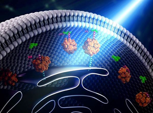
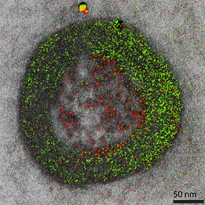
Illustration shows how “bridges” can be built within a cell through the use of light-reacting molecules. The light triggers proteins to line up within the cell, making it easier to shuttle molecules between the membranes of two subcellular organelles. This optogenetic strategy is helping scientists to control cell function with a simple beam of light. Illustration courtesy of Yubin Zhou.

Building Bridges

Yubin ZhouLink to external web site of Texas A&M is using optogenetics to control the way cells communicate and to study immune cell function. In one line of research, Zhou is using light to make it easier for calcium ions to enter cells. The ions carry instructions for the cell and also help tether small cellular structures (called organelles).  Those inter-membrane tethers allow for the movement of  proteins and lipids back and forth across the cell, and are critical for sending chemical messengers to communicate information (see illustration). When this process is disrupted, it can lead to extreme changes in cell function and even cell death. Using this technology to “switch on” normal pathways enables the scientists to better understand how such processes can be disrupted.

Continue reading “Optogenetics Sparks New Research Tools”

New Technology May Help Reduce Serious and Costly Post-Surgical Infections—Using Nothing but Air

7 comments

According to a recent estimate, implant infections following hip and knee replacement surgeries in the U.S. may number 65,000 by 2020, with the associated healthcare costs exceeding $1 billion. A new small, high-tech device could have a significant impact on improving health outcomes and reducing cost for these types of surgeries. The device, Air Barrier System (ABS), attaches on top of the surgical drape and gently emits HEPA-filtered air over the incision site. By creating a “cocoon” of clean air, the device prevents airborne particles—including the bacteria that can cause healthcare-associated infections—from entering the wound.

Air Barrier System
The Air Barrier System creates a “cocoon” of clean air (gray area with size indicated) over a surgical site to remove airborne contaminants and reduce the risk of infection in patients who are receiving an artificial hip, a blood vessel graft, a titanium plate in the spine or other implants.

Scientists recently analyzed the effectiveness of the ABS device in a clinical study—funded by NIGMS—involving nearly 300 patients. Each patient needed an implant, such as an artificial hip, a blood vessel graft in the leg or a titanium plate in the spine. Because implant operations involve inserting foreign materials permanently into the body, they present an even higher risk of infection than many other surgeries, and implant infections can cause life-long problems.

The researchers focused on one of the most common causes of implant infections—the air in the operating room. Although operating rooms are much cleaner than almost any other non-hospital setting, it’s nearly impossible to sterilize the entire room. Instead, the scientists focused on reducing contaminants directly over the surgical site. They theorized that if the air around the wound was cleaner, the number of implant infections might go down. Continue reading “New Technology May Help Reduce Serious and Costly Post-Surgical Infections—Using Nothing but Air”

Cool Tools: Pushing the Limits of High-Resolution Microscopy

1 comment

Cell biologists would love to shrink themselves down and actually see, touch and hear the inner workings of cells. Because that’s impossible, they have developed an ever-growing collection of microscopes to study cellular innards from the outside. Using these powerful tools, researchers can exhaustively inventory the molecular bits and pieces that make up cells, eavesdrop on cellular communication and spy on cells as they adapt to changing environments.

In recent years, scientists have developed new cellular imaging techniques that allow them to visualize samples in ways and at levels of detail never before possible. Many of these techniques build upon the power of electron microscopy (EM) to see ever smaller details.

Unlike traditional light microscopy, EM uses electrons, not light, to create an image. To do so, EM accelerates electrons in a vacuum, shoots them out of an electron gun and focuses them with doughnut-shaped magnets onto a sample. When electrons bombard the sample, some pass though without being absorbed while others are scattered. The transmitted electrons land on a detector and produce an image, just as light strikes a detector (or film) in a camera to create a photograph.

This image, showing a single protein molecule, is a montage. It was created to demonstrate how dramatically cryo-EM has improved in recent years. In the past, cryo-EM was only able to obtain a blobby approximation of a molecule’s shape, like that shown on the far left. Now, the technique yields exquisitely detailed images in which individual atoms are nearly visible (far right). Color is artificially applied. Credit: Veronica Falconieri, Subramaniam Lab, National Cancer Institute.

Transmission electron microscopes can magnify objects more than 10 million times, enabling scientists to see the outline and some details of cells, viruses and even some large molecules. A relatively new form of transmission electron microscopy called cryo-EM enables scientists to view specimens in their natural or near-natural state without the need for dyes or stains.

In cryo-EM—the prefix cry- means “cold” or “freezing”—scientists freeze a biological sample so rapidly that water molecules do not have time to form ice crystals, which could shove cellular materials out of their normal place. Cold samples are more stable and can be imaged many times over, allowing researchers to iteratively refine the image, remove artifacts and produce even sharper images than ever before. Continue reading “Cool Tools: Pushing the Limits of High-Resolution Microscopy”

Cool Image: Adding Color to the Gray World of Electron Microscopy

1 comment

Color electron micrograph of an endosome, a cell organelle. Credit: Ranjan Ramachandra, UCSD

As his Christmas gift to himself each year, the late biochemist Roger Tsien treated himself to two weeks of uninterrupted research in his lab. This image is a product of those annual sojourns. While it may look like a pine wreath dotted with crimson berries, it is in fact one of the world’s first color electron micrographs—and the method used to create it may dramatically advance cell imaging.

Electron microscopy (EM) is a time-honored technique for visualizing cell structures that uses beams of accelerated electrons to magnify objects up to 10 million times their actual size. Standard EM images are in grayscale and any color is added in with computer graphics programs after the image is made. With their new technique, Tsien, who received a Nobel Prize for his development of green fluorescent protein into a tool for visualizing details in living cells using light microscopes, and his colleagues have found a way to incorporate color labeling directly into EM. Continue reading “Cool Image: Adding Color to the Gray World of Electron Microscopy”

Visualizing Skin Regeneration in Real Time

2 comments
Top: Colorful skin cells on a zebrafish . Bottom: Cells from the outer surface of the scale.
More than 70 Skinbow colors distinguish hundreds of live cells from a tiny bit (0.0003348 square inches) of skin on the tail fin of an adult zebrafish. The bottom image shows the cells on the outer surface of a scale. Credit: Chen-Hui Chen, Duke University.

Zebrafish, blue-and-white-striped fish that are about 1.5 inches long, can regrow injured or lost fins. This feature makes the small fish a useful model organism for scientists who study tissue regeneration.

To better understand how zebrafish skin recovers after a scrape or amputation, researchers led by Kenneth Poss of Duke University tracked thousands of skin cells in real time. They found that lifespans of individual skin cells on the surface were 8 to 9 days on average and that the entire skin surface turned over in 20 days.

The scientists used an imaging technique they developed called “Skinbow,” which essentially shows the fish’s outer layer of skin cells in a spectrum of colors when viewed under a microscope. Skinbow is based on a technique created to study nerve cells in mice, another model organism.

The research team’s color-coded experiments revealed several unexpected cellular responses during tissue repair and replacement. The scientists plan to incorporate additional imaging techniques to generate an even more detailed picture of the tissue regeneration process.

The NIH director showcased the Skinbow technique and these images on his blog, writing: “You can see more than 70 detectable Skinbow colors that make individual cells as visually distinct from one another as jellybeans in a jar.”

This work was funded in part by NIH under grant R01GM074057.

Another Piece to a Century-Old Evolutionary Puzzle

0 comments
After mating about 55,000 pairs of fruit flies and sifting through 333,000 daughter flies, a research team found six sons that each had mutations in the same gene that helped make two fruit fly species unique from each other. Credit: Jim Woolace, Fred Hutch News Service.

Nitin Phadnis and Harmit Malik set out to conduct an experiment that could solve a century-old evolutionary puzzle: How did two related fruit fly species arise from one? Years after they began their quest, they finally have an answer.

The existence of a gene that helps make each of these fruit fly species unique and separate from each other had been guessed at since 1940, following experiments decades earlier in which geneticists first noticed that the two types of flies, when mated, had only daughters—no sons.

Scientists had previously discovered two other genes involved in driving the fruit fly species apart, but they knew those two genes weren’t the full story.

Continue reading “Another Piece to a Century-Old Evolutionary Puzzle”

Recognition for CRISPR Gene-Editing Tool

3 comments

The CRISPR gene-editing tool was recognized today by Science magazine as its “breakthrough of the year.” We support a number of researchers working in this exciting area and have featured it on this blog. To learn more about this exceptionally promising new method, see below for our illustrated explanation of the CRISPR system and its possible applications.

How the CRISPR System Works

Illustration of CRISPR system

The CRISPR system has two components joined together: a finely tuned targeting device (a small strand of RNA programmed to look for a specific DNA sequence) and a strong cutting device (an enzyme called Cas9 that can cut through a double strand of DNA).

CRISPR system in a cell

Once inside a cell, the CRISPR system locates the DNA it is programmed to find. The CRISPR seeking device recognizes and binds to the target DNA (circled, black).

The Cas9 enzyme cuts both strands of the DNA.

New genetic material incorporated into the broken DNA

Researchers can insert into the cell new sections of DNA. The cell automatically incorporates the new DNA into the gap when it repairs the broken DNA.

CRISPR has many possible uses, including:

  • Insert a new gene so the organism produces useful medicines.
  • Help treat genetic diseases.
  • Create tailor-made organisms to study human diseases.
  • Help produce replacements for damaged or diseased tissues and organs.

Cracking a Ubiquitous Code

0 comments

We asked the heads of our scientific divisions to tell us about some of the big questions in fundamental biomedical science that researchers are investigating with NIGMS support. This article is the third in an occasional series that explores these questions and explains how pursuing the answers could advance understanding of important biological processes.

Ubiquitin (Ub) molecules
Ubiquitin (Ub) molecules attached to proteins can form possibly hundreds of different shapes. Credit: NIGMS.

Researchers are on a quest to crack a code made by ubiquitin, a small protein that plays a big role in coordinating cellular function. By attaching to other proteins, ubiquitin determines what those proteins should do next.

Just as zip codes direct letters to specific towns, the ubiquitin code might direct one protein to help with DNA repair, another to assist in cell division, and a third to transport molecules into and out of cells. Continue reading “Cracking a Ubiquitous Code”