Category: Tools and Techniques

Interview With a Scientist – Rommie Amaro: Computational and Theoretical Model Builder

0 comments

Many researchers who search for anti-cancer drugs have labs filled with chemicals and tissue samples. Not Rommie Amaro. Her work uses computers to analyze the shape and behavior of a protein called p53. Defective versions of p53 are associated with more human cancers than any other malfunctioning protein.

Continue reading “Interview With a Scientist – Rommie Amaro: Computational and Theoretical Model Builder”

Teens Explore Science and Health through Game Design

0 comments

Educators often struggle to teach teens about sexual and reproductive health. Hexacago Health Academy (HHA) Link to external web site, an education program from the University of Chicago, leverages the fun activity of gameplay to impart these lessons to young people from Chicago’s South Side community. Funded by the Student Education Partnership Award (SEPA)Link to external web site, part of the National Institute of General Medical Sciences (NIGMS), in 2015, HHA assists teachers in their goal of helping teen students gain awareness and control over their health and also learn about careers in STEM Link to external web site and health fields.

Woman in a black buisness suit with arms crossed standing against a wall and smiling
Melissa Gilliam, founder of Ci3. Credit: Anna Knott, Chicago Magazine.

Genesis of HHA

HHA was cofounded by Melissa Gilliam Link to external web site, a University of Chicago professor of Obstetrics/Gynecology and Pediatrics and founder of the Center for Interdisciplinary Inquiry & Innovation in Sexual and Reproductive Health (Ci3) Link to external web site. During a 2013 summer program with high school students, Gilliam and Patrick Jagoda Link to external web site, associate professor of English and Cinema & Media Studies, and cofounder of Ci3’s Game Changer Chicago Design Lab, introduced the students to their STEM-based alternate reality game called The Source, in which a young woman crowdsources player help to solve a mystery that her father has created for her.

From their experience with The Source, Gilliam and Jagoda quickly learned that students not only wanted to play games but to design them too. What followed was the Game Changer Lab’s creation of the Hexacago game board, as well as the launch of HHA, a SEPA-funded project that the lab oversees.

Hexacago Game Board

At the core of HHA is the Hexacago game board, which displays the city of Chicago, along with Lake Michigan, a train line running through the city, and neighborhoods gridded into a hexagonal pattern.

HHA students not only play games designed from the Hexacago board template, but also design their own games from it that are intended to inspire behavior change in health-related situations and improve academic performance.

High school students seated at a table with a glossy, laminate test model of the Hexacago game and game pieces on top of it
Credit: Ci3 at the University of Chicago.

In this way, HHA is much more than just game design and play. “Students have no idea that what they’re doing is learning. In their minds, they’re really focused on designing games,” says Gilliam. “That’s the idea behind Hexacago Health Academy: helping people acquire deep knowledge of science and health issues by putting on the hat of a game designer.” Moreover, through the process of gameplay and design, students practice all the rich skills that result from teamwork, including collaborative learning, leadership, and communication.

Continue reading “Teens Explore Science and Health through Game Design”

Computational Geneticist Discusses Genetics of Storytelling at Sundance Film Festival

0 comments

About 10 years ago, University of Utah geneticist Mark Yandell developed a software platform called VAAST (Variant Annotation, Analysis & Search Tool) to identify rare genes. VAAST, which was funded by NHGRI, was instrumental in pinpointing the genetic cause of a mystery disease that killed four boys across two generations in an Ogden, UT family.

NIGMS has been supporting Yandell’s creation of the next generation of his software, called VAAST 2, for the past few years. The new version incorporates models of how genetic sequences are conserved among different species to improve accuracy with which benign genetic sequences can be differentiated from disease-causing variations. These improvements can help identify novel disease-causing genes responsible for both rare and common diseases.

Yandell and his colleagues in the Utah Genome Project recently took part in a panel at the Sundance Film Festival called the “Genetics of Storytelling” to discuss film’s ability to convey the power of science and medicine. Yandell told the audience his story about his efforts to use VAAST to trace the Ogden boys’ genetic variation back to their great-great-great-great-great grandmother.

Continue reading “Computational Geneticist Discusses Genetics of Storytelling at Sundance Film Festival”

What Zombie Ants Are Teaching Us About Fungal Infections: Q & A with Entomologists David Hughes and Maridel Fredericksen

0 comments

I can still remember that giddy feeling I had seven years ago, when I first read about the “zombie ant.” The story was gruesome and fascinating, and it was everywhere. Even friends and family who aren’t so interested in science knew the basics: in a tropical forest somewhere there’s a fungus that infects an ant and somehow takes control of the ant’s brain, forcing it to leave its colony, crawl up a big leaf, bite down and wait for the sweet relief of death. A grotesque stalk then sprouts from the poor creature’s head, from which fungal spores rain down to infect a new batch of ants.

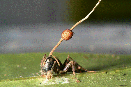
A fungal fruiting body erupts through the head of a carpenter ant infected by a parasitic fungus in Thailand. Credit: David Hughes, Penn State University.

The problem is, it doesn’t happen quite like that. David Hughes, the Penn State University entomologist who reported his extensive field observations of the fungus/ant interactions in BMC Ecology Exit icon, which caused much excitement back in 2011, has continued to study the fungus, Ophiocordyceps unliateralis, and its carpenter ant host, Camponotus leonardi.

In late 2017, Hughes and his colleagues published an article in PNAS Exit icon in which they used sophisticated microscopy and image-processing techniques to describe in great detail how the fungus invades various parts of the ant’s body including muscles in its legs and head.

Although Hughes’s earlier BMC Ecology paper showed fungus in the head of an ant, the new study reveals that the fungus never actually enters the brain.

To me, the new finding somehow made the fungus’ control over the ant even more baffling. What exactly was going on?

To find out, I spoke with Hughes and his graduate student Maridel Fredericksen.

Continue reading “What Zombie Ants Are Teaching Us About Fungal Infections: Q & A with Entomologists David Hughes and Maridel Fredericksen”

Fall 2017 Issue of Findings Magazine

0 comments

It’s back! Check out the new issue of Findings magazine.

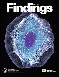
Findings Magazine Cover for Fall 2017.Findings presents cutting-edge research from scientists in diverse biomedical fields. The articles are aimed at high school students with the goal of making science—and the people who do it—interesting and exciting, and to inspire young readers to pursue careers in biomedical research. In addition to putting a face on science, Findings offers activities such as quizzes and crossword puzzles and, in its online version, video interviews with scientists.

The Fall 2017 issue profiles Yale University biologist Enrique De La Cruz, who studies how actin—a protein chain that supports cell structure—breaks so easily. Also profiled is University of California, Berkeley, biologist Rebecca Heald and her study of developmental factors that control an animal’s size.

This issue also features:

  • A virtual reality program designed to help burn patients manage pain
  • The promise of gene therapy for glaucoma
  • The many ways scientists categorize the biological world using “omics”
  • What researchers know—and don’t know—about how general anesthetics work
  • How animation helps researchers visualize interactions between biological molecules
  • How cells use sugary outer coatings to distinguish friend from foe
  • What makes our tissues stiff, squishy, solid, or see-through (hint: its initials are ECM)
  • How super-powerful microscopes are revealing views of biology never possible before

View Findings online, or order a print copy (classroom sets of up to 30 copies are available for educators).

Happy Birthday, BioBeat

0 comments

This month, our blog that highlights NIGMS-funded research turns four years old! For each candle, we thought we’d illuminate an aspect of the blog to offer you, our reader, an insider’s view.

Who are we?

Over the years, the editorial team has included onsite science writers, office interns, staff scientists and guest authors from universities. Kathryn, who’s a regular contributor, writes entirely from her home office. Chris, who has a Ph.D. in neuroscience and now manages the blog, used to do research in a lab. Alisa has worked in NIGMS’ Bethesda-based office the longest: 22 years! She and I remember when we first launched Biomedical Beat as an e-newsletter in 2005. You can read more about each of the writers on the contributors page and if you know someone who’s considering a career in science communications, tell them to drop us a line.

How do we come up with the stories?

We get our story ideas from a range of sources. For instance, newspaper articles about an experimental pest control strategy in Florida and California prompted us to write about NIGMS-funded studies exploring the basic science of the technique. A beautiful visual from a grantee’s institution inspired a short post on tissue regeneration research. And an ongoing conversation with NIGMS scientific staff about the important role of research organisms in biological studies sparked the idea for a playful profile of one such science superstar.

A big change in our storytelling has been shifting the focus from a single finding to broader progress in a lab or field. So instead of reporting on a study just published in a scientific journal, we may write about the scientist’s career path or showcase a collection of recent findings in that particular field. These approaches help us demonstrate that scientific understanding usually progresses through the slow and steady work undertaken by many labs.

What are our favorite posts?

I polled the writers on posts they liked, and the list is really long! Here are the top picks.


Four Ways Inheritance Is More Complex Than Mendel Knew
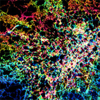

The Endoplasmic Reticulum: Networking in the Cell


Interview With a Scientist: Janet Iwasa, Molecular Animator


From Basic Research to Bioelectric Medicine
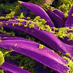

An Insider’s Look at Life: Magnified, an Airport Exhibit of Stunning Microscopy Images

What are your favorite posts?

We regularly review data about the number of times a blog post has been viewed to identify the ones that interest readers the most. That information also helps guide our decisions about other topics to feature on the blog. The Cool Image posts are among the most popular! Below are some other chart-topping posts.


Our Complicated Relationship With Viruses
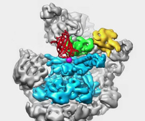

The Proteasome: The Cells Trash Processor in Action
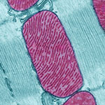

Demystifying General Anesthetics


Meet Sarkis Mazmanian and the Bacteria That Keep Us Healthy


5 Reasons Biologists Love Math

We always like hearing from readers! If there’s a basic biomedical research topic you’d like us to write about, or if you have feedback on a story or the blog in general, please leave your suggestions in the comment field below.

On Pi Day, Computational Biologists Share What They Love About Math

0 comments
Another cool fact about Pi: The mirror reflection of the numbers 3 1 4 spells out P I E.

Why do math lovers around the world call March 14 “Pi Day”? Because Pi, the ratio of a circle’s circumference to its diameter, is 3.14. Pi is a Greek letter (π) that represents a constant in math: All circles have the same Pi, regardless of their size. Pi has been calculated out to as many as 1 trillion digits past the decimal, and it can continue forever without repetition or pattern.

In honor of Pi Day, we asked several biomedical researchers in the field of computational biology to tell us why they love math and how they use it in their research. Continue reading “On Pi Day, Computational Biologists Share What They Love About Math”

Online Virus Tracking Tool Nextstrain Wins Inaugural Open Science Prize

0 comments
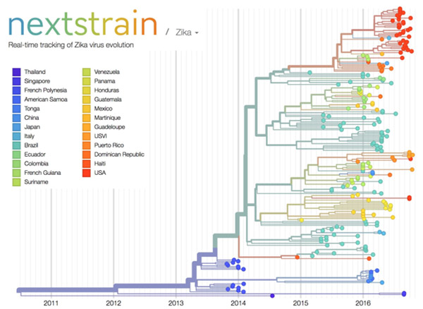
Nextstrain’s analysis of the genomes from Zika virus obtained in 25 countries over the past few years. 
Credit: Trevor Bedford and Richard Neher, nextstrain.org.

Over the past decade, scientists and clinicians have eagerly deposited their burgeoning biomedical data into publicly accessible databases. However, a lack of computational tools for sharing and synthesizing the data has prevented this wealth of information from being fully utilized.

In an attempt to unleash the power of open-access data, the National Institutes of Health, in collaboration with the Howard Hughes Medical Institute and Britain’s Wellcome Trust, launched the Open Science Prize. Last week, after a multi-stage public voting process, the inaugural award was announced. The winner of the grand prize—and $230,000—is a prototype computational tool called nextstrain Exit icon that tracks the spread of emerging viruses such as Ebola and Zika. This tool could be especially valuable in revealing the transmission patterns and geographic spread of new outbreaks before vaccines are available, such as during the 2013-2016 Ebola epidemic and the current Zika epidemic.

An international team of scientists—led by NIGMS grantee Trevor Bedford of the Fred Hutchinson Cancer Research Center, Seattle, and Richard Neher Exit icon of Biozentrum at the University of Basel, Switzerland—developed nextstrain as an open-access system capable of sharing and analyzing viral genomes. The system mines viral genome sequence data that researchers have made publicly available online. nextstrain then rapidly determines the evolutionary relationships among all the viruses in its database and displays the results of its analyses on an interactive public website.

The image here shows nextstrain’s analysis of the genomes from Zika virus obtained in 25 countries over the past few years. Plotting the relatedness of these viral strains on a timeline provides investigators a sense of how the virus has spread and evolved, and which strains are genetically similar. Researchers can upload genome sequences of newly discovered viral strains—in this case Zika—and find out in short order how their new strain relates to previously discovered strains, which could potentially impact treatment decisions.

Nearly 100 interdisciplinary teams comprising 450 innovators from 45 nations competed for the Open Science Prize. More than 3,500 people from six continents voted online for the winner. Other finalists for the prize focused on brain maps, gene discovery, air-quality monitoring, neuroimaging and drug discovery.

nextstrain was funded in part by NIH under grant U54GM111274.

Student Researcher Finds New Clues About Flu with Old Data

1 comment

Do you like to find new uses for old things? Like weaving old shirts into a rug, repurposing bottles into candle holders or turning packing crates into tables? Katie Gostic, a University of California, Los Angeles (UCLA) graduate student, likes finding new uses for old data. She channeled this interest when she analyzed existing data to study whether childhood exposure to flu affects a person’s future immunity to the disease.

Katie Gostic
Gostic conducted research for the flu project during the summer of 2015 when she was visiting her boyfriend, a tropical biologist, in Alamos, Sonora, Mexico. Credit: Charlie de la Rosa.

As an undergraduate student at Princeton University, Gostic was originally pursuing a degree in engineering. Her focus shifted to biology after taking an infectious disease modeling class. Gostic’s background in math and programming allows her to take large, complex pre-existing data sets and reanalyze them using new tools and methods. The result: Information that wasn’t accessible when the data were first collected.

Now a graduate researcher in the ecology and evolutionary biology lab of James Lloyd-Smith Exit icon, Gostic studies infectious diseases. The lab builds mathematical models to investigate zoonotic diseases—diseases that animals can transmit to humans but that humans don’t frequently spread between each other. Examples include diseases caused by Leptospira, a type of bacteria that infects household pets and many other animals, and monkeypox, a virus whose transmission to humans is increasing since the eradication of smallpox. The lab also studies bird flus, a category of flu viruses that infect birds and other animals and only occasionally jump to people. A very small number of cases of human-to-human transmission of bird flus have been recorded. However, if a bird flu virus mutated in a way that allowed it to spread among humans, it could cause a pandemic. Continue reading “Student Researcher Finds New Clues About Flu with Old Data”

Viral Views: New Insights on Infection Strategies

0 comments

The following images show a few ways in which cutting-edge research tools are giving us clearer views of viruses—and possible ways to disarm them. The examples, which highlight work involving HIV and the coronavirus, were funded in part by our Biomedical Technology Research Resources program.

Uncloaking HIV’s Camouflage

HIV capsid with (right, red) and without (left) a camouflaging human protein.
HIV capsid with (right, red) and without (left) a camouflaging human protein. Credit: Juan R. Perilla, Klaus Schulten and the Theoretical and Computational Biophysics Group, University of Illinois at Urbana-Champaign.

To sneak past our immune defenses and infect human cells, HIV uses a time-honored strategy—disguise. The virus’ genome is enclosed in a protein shell called a capsid (on left) that’s easily recognized and destroyed by the human immune system. To evade this fate, the chrysalis-shaped capsid cloaks itself with a human protein known as cyclophilin A (in red, on right). Camouflaged as human, the virus gains safe passage into and through a human cell to deposit its genetic material in the nucleus and start taking control of cellular machinery.

Biomedical and technical experts teamed up to generate these HIV models at near-atomic resolution. First, structural biologists at the Pittsburgh Center for HIV Protein Interactions Exit icon used a technique called cryo-electron microscopy (cryo-EM) to get information on the shape of an HIV capsid as well as the capsid-forming proteins’ connections to each other and to cyclophilin A. Then experts at the Resource for Macromolecular Modeling and Bioinformatics fed the cryo-EM data into their visualization and simulation programs to computationally model the physical interactions among every single atom of the capsid and the cyclophilin A protein. The work revealed a previously unknown site where cyclophilin A binds to the capsid, offering new insights on the biology of HIV infection. Continue reading “Viral Views: New Insights on Infection Strategies”