
Over the year, we dove into the inner workings of cells, interviewed award-winning researchers supported by NIGMS, shared a cool collection of science-themed backgrounds for video calls, and more. Here, we highlight three of the most popular posts from 2020. Tell us which of this year’s posts you liked best in the comments section below!
The Science of Infectious Disease Modeling
 Spike proteins on the surface of a coronavirus. Credit: David Veesler, University of Washington.
What does “modeling the spread” (or “flattening the curve”) mean, and how does it apply to infectious diseases such as COVID-19? Learn about the science of infectious disease modeling and how NIGMS supports scientists in the field.
Continue reading “Year in Review: Our Top Three Posts of 2020”
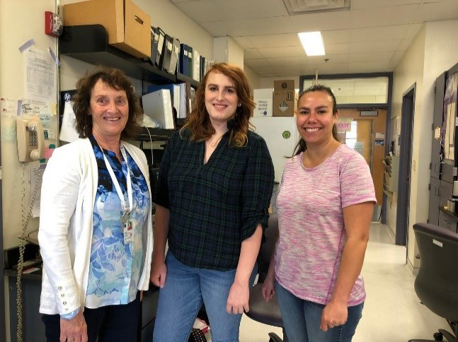 Dr. Wandinger-Ness (left) with former undergraduate trainee Amber Rauch (center) and current Ph.D. trainee Melanie Rivera. Credit: Angela Wandinger-Ness, Ph.D.
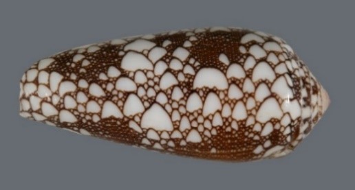 A cone snail shell. Credit: Kerry Matz, University of Utah.
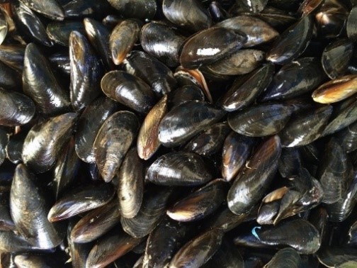 Mimicking mussels’ natural “glue” could have multiple benefits.
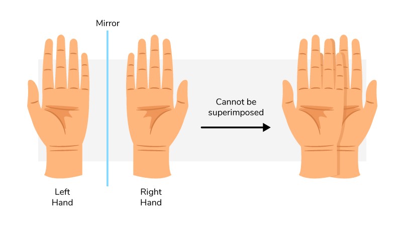 Our hands are chiral: They’re mirror images but aren’t identical.