Category: Tools and Techniques

Quiz: What Can Research Organisms Reveal About Health?

0 comments

Scientists often use research organisms to study life. Examples range from simple organisms like bacteria to more complex ones such as mice. NIGMS funds studies of research organisms to understand biological processes that are common to all organisms, including humans. Errors in these fundamental processes can cause disease, and better understanding of these malfunctions can aid in the development of potential treatments.

Research organisms may also reveal novel biological processes that can lead to important scientific or medical technologies. For example, researchers studying interactions between viruses and bacteria made a discovery that led to the CRISPR (clustered regularly interspaced short palindromic repeats) gene-editing system, which was recognized by the 2020 Nobel Prize in chemistry.

Continue reading “Quiz: What Can Research Organisms Reveal About Health?”

Fifty Years of the Protein Data Bank!

0 comments
Macromolecular structures in the shape of the number 50. Protein Data Bank’s 50 years logo. Credit: PDB website.

The Protein Data Bank (PDB), established in 1971, is the single global repository for 3D structural data of proteins, DNA, RNA, and even complexes these biological molecules form with drugs or other small molecules. More than 1 million people—including researchers, medical professionals, educators, and students—use the PDB each year. NIGMS and other parts of NIH have helped fund this free digital resource since 1978.

Continue reading “Fifty Years of the Protein Data Bank!”

Biology Beyond the Lab: Using Computers to Study Life

1 comment
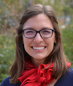
A headshot of Dr. Melissa Wilson.
Learn more about Dr. Melissa Wilson’s computational biology research in another Biomedical Beat blog post. Credit: Jacob Sahertian, ASU.

“You’re not going to be able to do biology without understanding programming in the future,” Melissa Wilson, Ph.D., an associate professor of genomics, evolution, and bioinformatics at Arizona State University, said in her 2019 NIGMS Early Career Investigator Lecture. “You don’t have to be an expert programmer. But without understanding programming, I can assert you won’t be able to do biology in the next 20 years.”

A growing number of researchers, like Dr. Wilson, are studying biology using computers and mathematical methods. Some of them started in traditional biology or other life science labs, while others studied computer science or math first. Here, we’re featuring two researchers who took different paths to computational biology.

Continue reading “Biology Beyond the Lab: Using Computers to Study Life”

A Focus on Microscopes: See Eye-Catching Images

3 comments

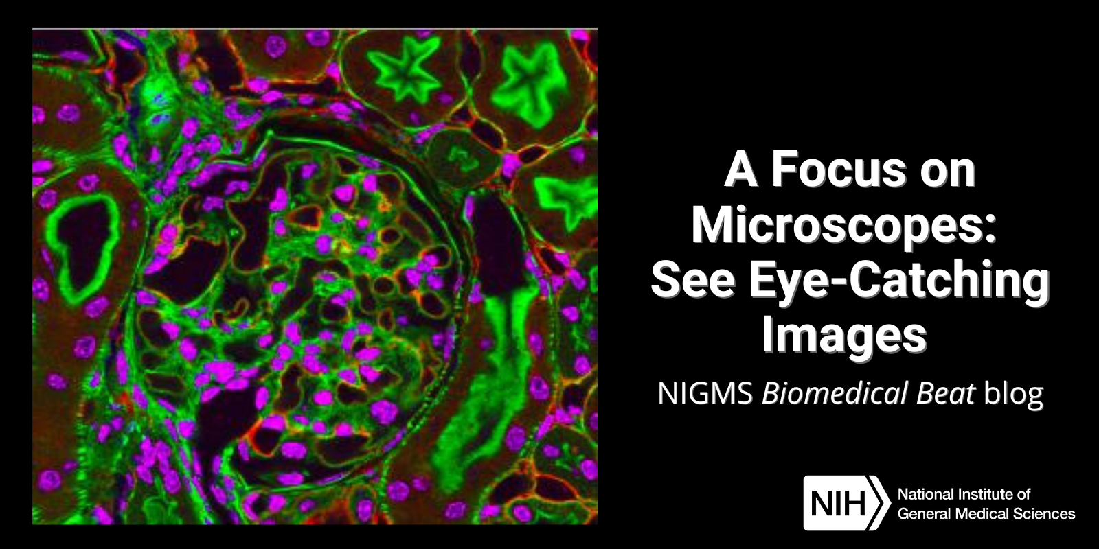
Have you ever wondered what creates striking images of cells and other tiny structures? Most often, the answer is microscopes. Many of us have encountered basic light microscopes in science classes, but those are just one of many types that scientists use. Check out the slideshow to see images researchers have captured using different kinds of microscopes. For even more images of the microscopic world, visit the NIGMS Image and Video Gallery.

Continue reading “A Focus on Microscopes: See Eye-Catching Images”

Quiz: Are You a Genetics Genius?

3 comments

Genes are segments of DNA. They contain instructions for building one or more molecules that help the body work. Researchers in the field of genetics study genes and heredity—how certain traits are passed from parents to their offspring through DNA. NIGMS supports many scientists who investigate the genetics of people and research organisms to better understand human health and disease.

Take our quiz below to test how much you know about genetics. For more quizzes and other fun learning tools, visit our activities and multimedia webpage.

Continue reading “Quiz: Are You a Genetics Genius?”

Pathways: The Imaging Issue

0 comments
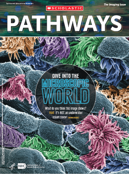
Cover of Pathways student magazine showing geometric shapes, pom-pom-like structures, and text that reads, Dive into the microscopic world. What do you think this image shows? Hint: It’s NOT an underwater scene! (Answer inside). Cover of Pathways student magazine.

NIGMS and Scholastic bring you our latest issue of Pathways, which focuses on imaging tools that help scientists unlock the mysteries of our cells and molecules. A better understanding of this tiny world can help researchers learn about the body’s normal and abnormal processes and lead to more effective, targeted treatments for illnesses.

Pathways is designed for students in grades 6 through 12. This collection of free resources teaches students about basic science and its importance to health, as well as exciting research careers.

Continue readingPathways: The Imaging Issue”

Year in Review: Our Top Three Posts of 2020

0 comments

Over the year, we dove into the inner workings of cells, interviewed award-winning researchers supported by NIGMS, shared a cool collection of science-themed backgrounds for video calls, and more. Here, we highlight three of the most popular posts from 2020. Tell us which of this year’s posts you liked best in the comments section below!

The Science of Infectious Disease Modeling

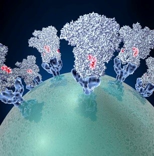
Oblong light-blue structures with red spots in the middle connected to the surface of a sphere. Spike proteins on the surface of a coronavirus. Credit: David Veesler, University of Washington.

What does “modeling the spread” (or “flattening the curve”) mean, and how does it apply to infectious diseases such as COVID-19? Learn about the science of infectious disease modeling and how NIGMS supports scientists in the field.

Continue reading “Year in Review: Our Top Three Posts of 2020”

An Enlightening Protein

0 comments
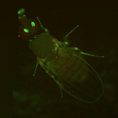
A fly glowing green. A fruit fly expressing GFP. Credit: Jay Hirsh, University of Virginia.

During the holiday season, twinkling lights are a common sight. But no matter what time of the year, you can see colorful glows in many biology labs. Scientists have enabled many organisms to light up in the dark—from cells to fruit flies and Mexican salamanders. These glowing organisms help researchers better understand basic cell processes because their DNA has been edited to express molecules such as green fluorescent protein.

Continue reading “An Enlightening Protein”

Q&A With Nobel Laureate and CRISPR Scientist Jennifer Doudna

0 comments
A headshot of Dr. Doudna. Jennifer Doudna, Ph.D. Credit: University of California, Berkeley.

The 2020 Nobel Prize in Chemistry was awarded to Jennifer Doudna, Ph.D., and Emmanuelle Charpentier, Ph.D., for the development of the gene-editing tool CRISPR. Dr. Doudna shared her thoughts on the award and answered questions about CRISPR in a live chat with NIH Director Francis S. Collins, M.D., Ph.D. Here are a few highlights from the interview.

Q: How did you find out that you won the Nobel Prize?

A: It’s a little bit of an embarrassing story. I slept through a very important phone call and finally woke up when a reporter called me. I was just coming out of a deep sleep, and the reporter was asking, “What do you think about the Nobel?” And I said, “I don’t know anything about it. Who won it?” I thought they were asking for comments on somebody else who won it. And she said, “Oh my gosh! You don’t know! You won it!”

Continue reading “Q&A With Nobel Laureate and CRISPR Scientist Jennifer Doudna”

Freezing a Moment in Time: Snapshots of Cryo-EM Research

0 comments

To get a look at cell components that are too small to see with a normal light microscope, scientists often use cryo-electron microscopy (cryo-EM). As the prefix cryo- means “cold” or “freezing,” cryo-EM involves rapidly freezing a cell, virus, molecular complex, or other structure to prevent water molecules from forming crystals. This preserves the sample in its natural state and keeps it still so that it can be imaged with an electron microscope, which uses beams of electrons instead of light. Some electrons are scattered by the sample, while others pass through it and through magnetic lenses to land on a detector and form an image.

Typically, samples contain many copies of the object a scientist wants to study, frozen in a range of orientations. Researchers take images of these various positions and combine them into a detailed 3D model of the structure. Electron microscopes allow us to see much smaller structures than light microscopes do because the wavelengths of electrons are much shorter than the wavelength of light. NIGMS-funded researchers are using cryo-EM to investigate a range of scientific questions.

Caught in Translation
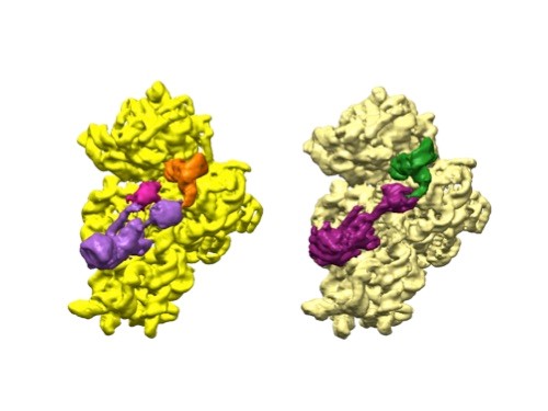
One cluster that is yellow, purple, and orange and another that is beige, purple, and green. 3D reconstructions of two stages in the assembly of the bacterial ribosome created from time-resolved cryo-EM images. Credit: Joachim Frank, Columbia University.

Joachim Frank, Ph.D., a professor of biochemistry and molecular biophysics and of biological sciences at Columbia University in New York, New York, along with two other researchers, won the 2017 Nobel Prize in Chemistry for developing cryo.

Dr. Frank’s lab focuses on the process of translation, where structures called ribosomes turn genetic instructions into proteins, which are needed for many chemical reactions that support life. Recently, Dr. Frank has adopted and further developed a technique called time-resolved cryo-EM. This method captures images of short-lived states in translation that disappear too quickly (after less than a second) for standard cryo-EM to capture. The ability to fully visualize translation could help researchers identify errors in the process that lead to disease and also to develop treatments.

Continue reading “Freezing a Moment in Time: Snapshots of Cryo-EM Research”