Tag: Research Organisms

Cool Images: Wondrous Worms

0 comments

The tiny roundworm Caenorhabditis elegans is one of the most common research organisms—creatures scientists use to study life. While C. elegans may seem drastically different from humans, it shares many genes and molecular pathways with us. Viewed with a microscope, the worm can also be surprisingly beautiful. Aside from the stunning imagery, these examples from our Image and Video Gallery show how C. elegans helps scientists advance our understanding of living systems and find new ways to improve our health.

Round yellow shapes with smaller blue spots. Three of the yellow shapes are connected by a purple line. 
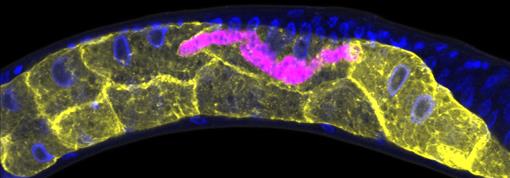
Credit: Keir Balla and Emily Troemel, University of California San Diego.

This C. elegans has been infected with microsporidia (purple), parasites closely related to fungi. The yellow shapes are the worm’s gut cells, and the blue dots are nuclei. Some microsporidia can infect people, so studying the parasites in worms could help researchers devise strategies to prevent or treat infections.

Continue reading “Cool Images: Wondrous Worms”

Reusable Disinfectant Developed from Mussel “Glue”

0 comments
A pile of ocean mussels with shiny black shells.
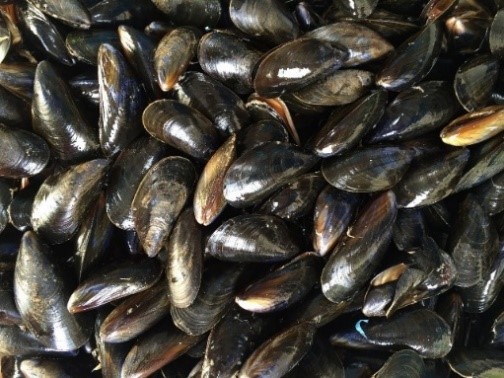
Mimicking mussels’ natural “glue” could have multiple benefits.

Many species have developed unique adaptations to help them thrive in their environments, and scientists in a field called biomimicry use these examples as the basis for tools to help humans. Biomimicry researchers have made a wide range of products, from climbing pads modeled after gecko feet to a faster, sharp-nosed bullet train based on the beak of the kingfisher bird. The animal kingdom also provides inspiration for biomedical products. For instance, scientists at Michigan Technological University in Houghton discovered that a natural “glue” produced by mussels has antimicrobial properties and are developing a way to put these properties to use.

Continue reading “Reusable Disinfectant Developed from Mussel “Glue””

Looking Back at the Top Three Posts of 2019

0 comments

Over the past 12 months, we’ve explored a variety of topics in genetics, cell biology, chemistry, and careers in the biomedical sciences. As we ring in the new year, we bring you our top three posts of 2019. If your favorite is missing, let us know what it is in the comments section below!

Amazing Organisms and the Lessons They Can Teach Us

Two Hawaiian bobtail squid with yellow skin, brown spots, and black eyes catching a neon green reflection. 
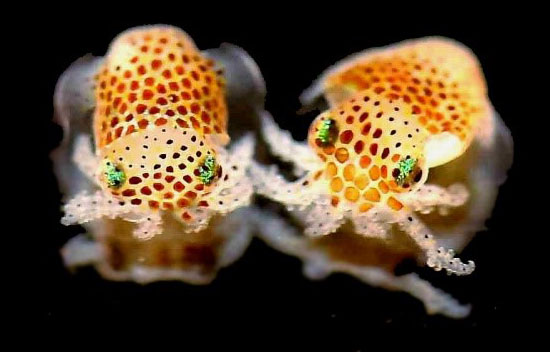
Hawaiian bobtail squid. Credit: Dr. Satoshi Shibata.

Studying research organisms, such as those featured in this post, teaches us about ourselves. These amazing creatures, which have some traits similar to our own, may hold the key to preventing and treating an array of complex diseases.

Continue reading “Looking Back at the Top Three Posts of 2019”

A Scientist’s Exploration of Regeneration

3 comments
Viravuth (“Voot”) Yin, standing with arms crossed and smiling in front of a shelves holding tanks of zebrafish in his lab. Viravuth (“Voot”) Yin, associate professor of regenerative biology and medicine at MDI Biological Laboratory and chief scientific officer at Novo Biosciences, Inc., in Bar Harbor, Maine. Credit: MDI Biological Laboratory.

In 1980, a week after his 6th birthday, Viravuth (“Voot”) Yin immigrated with his mother, grandfather, and three siblings from Cambodia to the United States. Everything they owned fit into a single, 18-inch carry-on bag. They had to build new lives from almost nothing. So, it’s perhaps fitting that Yin studies regeneration, the fascinating ability of some animals, such as salamanders, sea stars, and zebrafish, to regrow damaged body parts, essentially from scratch.

Yin’s path wasn’t always smooth. His family settled in Hartford, Connecticut, near an uncle who had been granted asylum during the Vietnam War. Yin got into a lot of trouble in school, trying to learn a new culture and fit in. Things improved when his mother moved him and his siblings to West Hartford, well known for its strong schools.

Continue reading “A Scientist’s Exploration of Regeneration”

Amazing Organisms and the Lessons They Can Teach Us

1 comment

What do you have in common with rodents, birds, and reptiles? A lot more than you might think. These creatures have organs and body systems very similar to our own: a skeleton, digestive tract, brain, nervous system, heart, network of blood vessels, and more. Even so-called “simple” organisms such as insects and worms use essentially the same genetic and molecular pathways we do. Studying these organisms provides a deeper understanding of human biology in health and disease, and makes possible new ways to prevent, diagnose, and treat a wide range of conditions.

Historically, scientists have relied on a few key organisms, including bacteria, fruit flies, rats, and mice, to study the basic life processes that run bodily functions. In recent years, scientists have begun to add other organisms to their toolkits. Many of these newer research organisms are particularly well suited for a specific type of investigation. For example, the small, freshwater zebrafish grows quickly and has transparent embryos and see-through eggs, making it ideal for examining how organs develop. Organisms such as flatworms, salamanders, and sea urchins can regrow whole limbs, suggesting they hold clues about how to improve wound healing and tissue regeneration in humans.

Continue reading “Amazing Organisms and the Lessons They Can Teach Us”

Interview with a Scientist: Jeramiah Smith on the Genomic Antics of an Ancient Vertebrate

0 comments

The first known descriptions of cancer come from ancient Egypt more than 3,500 years ago. Early physicians attributed the disease to several factors, including an imbalance in the body’s humoral fluids, trauma, and parasites. Only in the past 50 years or so have we figured out that mutations in critical genes are often the trigger. The sea lamprey, a slimy, snake-like blood sucker, is proving to be an ideal tool for understanding these mutations.

The sea lamprey, often called the jawless fish, is an ancient vertebrate whose ancestor diverged from the other vertebrate lineages (fish, reptiles, birds and mammals) more than 500 million years ago. Jeramiah Smith,Link to external web site associate professor of biology at the University of Kentucky, has discovered that lamprey have two separate genomes: a complete genome specific to their reproductive cells, consisting of 99 chromosomes (humans have 23 pairs) and another genome in which about 20 percent of genes have been deleted after development. Using the lamprey model, Smith and his colleagues have learned that many of these deleted genes—such as those that initiate growth pathways—are similar to human oncogenes (i.e., cancer-causing genes).

Continue reading “Interview with a Scientist: Jeramiah Smith on the Genomic Antics of an Ancient Vertebrate”

Genomic Gymnastics of a Single-Celled Ciliate and How It Relates to Humans

0 comments
Laura Landweber
Credit: Denise Applewhite.
Laura Landweber
Grew up in: Princeton, New Jersey
Job site: Columbia University, New York City
Favorite food: Dark chocolate and dark leafy greens
Favorite music: 1940’s style big band jazz
Favorite hobby: Swing dancing
If I weren’t a scientist I would be a: Chocolatier (see “Experiments in Chocolate” sidebar at bottom of story)

One day last fall, molecular biologist Laura Landweber surveyed the Princeton University lab where she’d worked for 22 years. She and her team members had spent many hours that day laboriously affixing yellow Post-it notes to the laboratory equipment—microscopes, centrifuges, computers—they would bring with them to Columbia University, where Landweber had just been appointed full professor. Each Post-it specified the machinery’s location in the new lab. Items that would be left behind—glassware, chemical solutions, furniture, office supplies—were left unlabeled.

As Landweber viewed the lab, decorated with a field of sunny squares, her thoughts turned to another sorting process—the one used by her primary research subject, a microscopic organism, to sift through excess DNA following mating. Rather than using Post-it notes, the creature, a type of single-celled organism called a ciliate, uses small pieces of RNA to tag which bits of genetic material to keep and which to toss.

Landweber is particularly fond of Oxytricha trifallax, a ciliate with relatives that live in soil, ponds and oceans all over the world. The kidney-shaped cell is covered with hair-like projections called cilia that help it move around and devour bacteria and algae. Oxytricha is not only bizarre in appearance, it’s also genetically creative.

Unlike humans, whose cells are programmed to die rather than pass on genomic errors, Oxytricha cells appear to delight in genomic chaos. During sexual reproduction, the ciliate shatters the DNA in one of its two nuclei into hundreds of thousands of pieces, descrambles the DNA letters, throws most away, then recombines the rest to create a new genome.

Landweber has set out to understand how—and possibly why—Oxytricha performs these unusual genomic acrobatics. Ultimately, she hopes that learning how Oxytricha rearranges its genome can illuminate some of the events that go awry during cancer, a disease in which the genome often suffers significant reorganization and damage.

Oxytricha’s Unique Features

Oxytricha carries two separate nuclei—a macronucleus and a micronucleus. The macronucleus, by far the larger of the two, functions like a typical genome, the source of gene transcription for proteins. The tiny micronucleus only sees action occasionally, when Oxytricha reproduces sexually.

Oxytricha trifallax cells in the process of mating
Two Oxytricha trifallax cells in the process of mating. Credit, Robert Hammersmith.

What really makes Oxytricha stand out is what it does with its DNA during the rare occasions that it has sex. When food is readily available, Oxytricha procreates without a partner, like a plant grown from a cutting. But when food is scarce, or the cell is stressed, it seeks a mate. When two Oxytricha cells mate, the micronuclear genomes in each cell swap DNA, then replicate. One copy of the new hybrid micronucleus remains intact, while the other breaks its DNA into hundreds of thousands of pieces, some of which are tagged, recombined, then copied another thousand-fold to form a new macronucleus. Continue reading “Genomic Gymnastics of a Single-Celled Ciliate and How It Relates to Humans”

Carole LaBonne: Neural Crest Cells and the Rise of the Vertebrates

0 comments

The stunning pigmentation of tigers, the massive jaws of sharks, and the hyper-acute vision of eagles. These and other remarkable features of higher organisms (vertebrates) derive from a small group of powerful cells, called neural crest cells, that arose more than 500 million years ago. Molecular biologist Carole LaBonne Exit icon of Northwestern University in Illinois studies how neural crest cells help give rise to these important vertebrate structures throughout development.

Very early during embryonic development, stem cells differentiate into different layers: mesoderm, endoderm, and ectoderm. Each of these layers then gives rise to different cell and tissue types. For example, the ectoderm becomes skin and nerve cells. Mesoderm turns into muscle, bone, fat, blood and the circulatory system. Endoderm forms internal structures such as lungs and digestive organs.

These three layers are present in vertebrates—animals with a backbone and well-defined heads, such as fish, birds, reptiles, and mammals—as well as animals without backbones, such as the marine-dwelling Lancelets and Tunicates (referred to as non-vertebrate chordates). Unlike cells in these layers, neural crest cells, which are found only in vertebrates, don’t specialize until much later in development. The delay gives neural crests cells the extra time and flexibility to sculpt the complex anatomical structures found only in vertebrate animals.

Scientists have long debated how neural crest cells manage to finalize their destiny so much later than all other cell types.

Using the frog Xenopus as a model system, LaBonne and her colleagues performed a series of experiments that revealed the process and identified key genes that control it.

In this video, LaBonne describes the power of neural crest cells and how they can be useful for studies of human health, including how cancer cells can metastasize, or migrate, throughout the body.

Dr. LaBonne’s research is funded in part by NIGMS grant 5R01GM116538.

Zebrafish Scrapbook

4 comments
Name: Danio rerio
Hometown: Freshwater ponds and rivers of India, Nepal, and neighboring countries
Occupation: Research
Long-term goal: Solving the basic mysteries of life
Work site: More than 600 science labs worldwide
Danio rerio

That’s me and some other zebrafish, swimming in a tank in one of the more than 600 labs around the world that use us to study embryo development, genetics, and all kinds of human diseases. Credit: Wikimedia Commons, Azul.

Apart from the tell-tale stripes that give me my nickname, zebrafish, I look a lot like your standard minnow swimming in the shallows of any pond, lake, or river. But I like to think I’m more important than that. In fact, researchers around the world have turned to me and my extended family to understand some of the most basic mysteries of life. From studying us, they’re learning about how embryos develop, how cancer works, and whether someday humans might be able to rebuild a heart, repair a spinal cord injury, or regrow a severed limb.

Why us? Because zebrafish are pretty special and researchers think we’re easy to work with. First, unlike your standard lab mouse or rat, we lay lots of eggs, producing baby fish that grow up fast. We develop outside our mothers and go from egg to embryo to free-swimming larva in just 3 days (check out this video Exit icon of how we grow, cell by cell, during the first 24 hours). Within 3 months, we’re fully mature.

Not only do zebrafish moms have many babies at the same time, and not only do these babies grow up quickly, but our eggs and embryos are see-through, so scientists can literally watch us grow one cell at a time. We stay mostly transparent for a few weeks after hatching. That makes it super easy for scientists to monitor us for both normal and abnormal development. In fact, scientists have learned how to turn off the genes that give our skin its color. These zebrafish, named casper, after the “friendly ghost” of cartoon fame, stay semi-transparent, or translucent, through adulthood.

And last, but certainly not least, did I mention that we can regenerate? If parts of my body are damaged, even to a significant degree, they can regrow. This holds true for my heart, fins, spinal cord, and even brain tissue. Our regenerative capacity is seemingly unlimited; my caudal fin, for example, can grow back dozens of times. Continue reading “Zebrafish Scrapbook”