Author: Emily Carlson

Headshot of emily Carlson

Posts by Emily Carlson

Happy Birthday, BioBeat

0 comments

This month, our blog that highlights NIGMS-funded research turns four years old! For each candle, we thought we’d illuminate an aspect of the blog to offer you, our reader, an insider’s view.

Who are we?

Over the years, the editorial team has included onsite science writers, office interns, staff scientists and guest authors from universities. Kathryn, who’s a regular contributor, writes entirely from her home office. Chris, who has a Ph.D. in neuroscience and now manages the blog, used to do research in a lab. Alisa has worked in NIGMS’ Bethesda-based office the longest: 22 years! She and I remember when we first launched Biomedical Beat as an e-newsletter in 2005. You can read more about each of the writers on the contributors page and if you know someone who’s considering a career in science communications, tell them to drop us a line.

How do we come up with the stories?

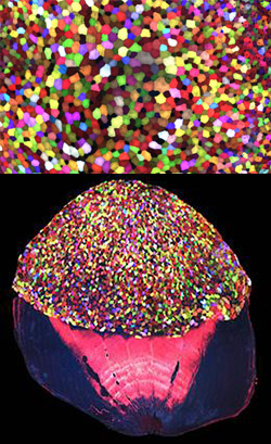
We get our story ideas from a range of sources. For instance, newspaper articles about an experimental pest control strategy in Florida and California prompted us to write about NIGMS-funded studies exploring the basic science of the technique. A beautiful visual from a grantee’s institution inspired a short post on tissue regeneration research. And an ongoing conversation with NIGMS scientific staff about the important role of research organisms in biological studies sparked the idea for a playful profile of one such science superstar.

A big change in our storytelling has been shifting the focus from a single finding to broader progress in a lab or field. So instead of reporting on a study just published in a scientific journal, we may write about the scientist’s career path or showcase a collection of recent findings in that particular field. These approaches help us demonstrate that scientific understanding usually progresses through the slow and steady work undertaken by many labs.

What are our favorite posts?

I polled the writers on posts they liked, and the list is really long! Here are the top picks.


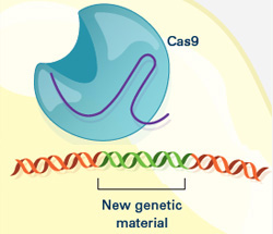
Four Ways Inheritance Is More Complex Than Mendel Knew


The Endoplasmic Reticulum: Networking in the Cell


Interview With a Scientist: Janet Iwasa, Molecular Animator


From Basic Research to Bioelectric Medicine


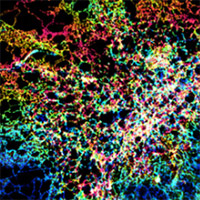
An Insider’s Look at Life: Magnified, an Airport Exhibit of Stunning Microscopy Images

What are your favorite posts?

We regularly review data about the number of times a blog post has been viewed to identify the ones that interest readers the most. That information also helps guide our decisions about other topics to feature on the blog. The Cool Image posts are among the most popular! Below are some other chart-topping posts.


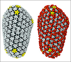
Our Complicated Relationship With Viruses


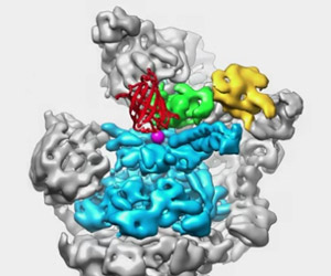
The Proteasome: The Cells Trash Processor in Action


Demystifying General Anesthetics


Meet Sarkis Mazmanian and the Bacteria That Keep Us Healthy


5 Reasons Biologists Love Math

We always like hearing from readers! If there’s a basic biomedical research topic you’d like us to write about, or if you have feedback on a story or the blog in general, please leave your suggestions in the comment field below.

Chasing Fireflies—and Better Cellular Imaging Techniques

0 comments
A glowing firefly sitting in a person's open palm. 
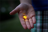
Firefly. Credit: Stock photo.

The yellow-green glow from this summer’s fireflies teased my kids across the yard. Max and Stella zigzagged the grass, occasionally jumping into the air to cup a firefly in their hands and then proudly shouting, “I got one!”

Chasing fireflies on a summer night is a childhood rite of passage for many, including Nathan Shaner who grew up in New Jersey. “It was one of my favorite things about summer,” he recalls. “I’d catch them with my hands—I’d never jar them.”

Today, Shaner studies the science of bioluminescence, which gives fireflies and many other organisms the natural ability to emit light. His goal is to make bright bioluminescent tags that he and other scientists can use to study living cells in greater detail. “There’s this very beautiful thing that evolved in nature, and we can use it to enable new discoveries,” he says.

Thousands of organisms glow as a way to communicate, spook predators, lure prey or attract mates. There are a few terrestrial examples, such as fireflies, glowworm insect larvae and foxfire fungi, and many more aquatic ones, including types of marine plankton, fish, jellyfish, shrimp, squid and sea urchins. One research team estimated nearly three quarters of sea life have bioluminescent capabilities.

Continue reading “Chasing Fireflies—and Better Cellular Imaging Techniques”

On Pi Day, Computational Biologists Share What They Love About Math

0 comments
Another cool fact about Pi: The mirror reflection of the numbers 3 1 4 spells out P I E.

Why do math lovers around the world call March 14 “Pi Day”? Because Pi, the ratio of a circle’s circumference to its diameter, is 3.14. Pi is a Greek letter (π) that represents a constant in math: All circles have the same Pi, regardless of their size. Pi has been calculated out to as many as 1 trillion digits past the decimal, and it can continue forever without repetition or pattern.

In honor of Pi Day, we asked several biomedical researchers in the field of computational biology to tell us why they love math and how they use it in their research. Continue reading “On Pi Day, Computational Biologists Share What They Love About Math”

Protein Paradox: Enrique De La Cruz Aims to Understand Actin

1 comment
Enrique M. De La Cruz
Credit: Jeff Foley, American Heart Association.
Enrique M. De La Cruz
Grew up in: Newark and Kearny, New Jersey
Job site: Yale University
Favorite food: His mom’s Spanish-style polenta (harina de maíz)
Alternative career: Managing a vinyl record shop
Favorite song: “Do Anything You Wanna Do” by Eddie & The Hot Rods

Enrique De La Cruz stood off to the side in a packed room. As he waited for his turn to speak, he stroked the beads of a necklace. Was he nervous? Quietly praying? When he took center stage, the purpose of the strand became clear.

Like a magician—and dressed all in black—De La Cruz held up the necklace with two hands so everyone, even those sitting in the back, could see it. It was made of snap-together beads. De La Cruz waved the strand. It wiggled in different directions. Then, with no sleight of hand, he popped off one of the beads. The necklace broke into two.

For the next hour, De La Cruz pulled out one prop after another: a piece of rope from his pocket, a pencil tucked behind his ear and even a fresh spear of asparagus stuffed in his backpack. At one point, De La Cruz assembled a conga line with people in the front row. Continue reading “Protein Paradox: Enrique De La Cruz Aims to Understand Actin”

Viral Views: New Insights on Infection Strategies

0 comments

The following images show a few ways in which cutting-edge research tools are giving us clearer views of viruses—and possible ways to disarm them. The examples, which highlight work involving HIV and the coronavirus, were funded in part by our Biomedical Technology Research Resources program.

Uncloaking HIV’s Camouflage

HIV capsid with (right, red) and without (left) a camouflaging human protein.
HIV capsid with (right, red) and without (left) a camouflaging human protein. Credit: Juan R. Perilla, Klaus Schulten and the Theoretical and Computational Biophysics Group, University of Illinois at Urbana-Champaign.

To sneak past our immune defenses and infect human cells, HIV uses a time-honored strategy—disguise. The virus’ genome is enclosed in a protein shell called a capsid (on left) that’s easily recognized and destroyed by the human immune system. To evade this fate, the chrysalis-shaped capsid cloaks itself with a human protein known as cyclophilin A (in red, on right). Camouflaged as human, the virus gains safe passage into and through a human cell to deposit its genetic material in the nucleus and start taking control of cellular machinery.

Biomedical and technical experts teamed up to generate these HIV models at near-atomic resolution. First, structural biologists at the Pittsburgh Center for HIV Protein Interactions Exit icon used a technique called cryo-electron microscopy (cryo-EM) to get information on the shape of an HIV capsid as well as the capsid-forming proteins’ connections to each other and to cyclophilin A. Then experts at the Resource for Macromolecular Modeling and Bioinformatics fed the cryo-EM data into their visualization and simulation programs to computationally model the physical interactions among every single atom of the capsid and the cyclophilin A protein. The work revealed a previously unknown site where cyclophilin A binds to the capsid, offering new insights on the biology of HIV infection. Continue reading “Viral Views: New Insights on Infection Strategies”

Visualizing Skin Regeneration in Real Time

2 comments
Top: Colorful skin cells on a zebrafish . Bottom: Cells from the outer surface of the scale.
More than 70 Skinbow colors distinguish hundreds of live cells from a tiny bit (0.0003348 square inches) of skin on the tail fin of an adult zebrafish. The bottom image shows the cells on the outer surface of a scale. Credit: Chen-Hui Chen, Duke University.

Zebrafish, blue-and-white-striped fish that are about 1.5 inches long, can regrow injured or lost fins. This feature makes the small fish a useful model organism for scientists who study tissue regeneration.

To better understand how zebrafish skin recovers after a scrape or amputation, researchers led by Kenneth Poss of Duke University tracked thousands of skin cells in real time. They found that lifespans of individual skin cells on the surface were 8 to 9 days on average and that the entire skin surface turned over in 20 days.

The scientists used an imaging technique they developed called “Skinbow,” which essentially shows the fish’s outer layer of skin cells in a spectrum of colors when viewed under a microscope. Skinbow is based on a technique created to study nerve cells in mice, another model organism.

The research team’s color-coded experiments revealed several unexpected cellular responses during tissue repair and replacement. The scientists plan to incorporate additional imaging techniques to generate an even more detailed picture of the tissue regeneration process.

The NIH director showcased the Skinbow technique and these images on his blog, writing: “You can see more than 70 detectable Skinbow colors that make individual cells as visually distinct from one another as jellybeans in a jar.”

This work was funded in part by NIH under grant R01GM074057.

The Proteasome: The Cell’s Trash Processor in Action

2 comments

Our cells are constantly removing and recycling molecular waste. On the occasion of Earth Day, we put together this narrated animation to show you one way cells process their trash. The video features the proteasome, a cellular machine that breaks down damaged or unwanted proteins into bits that the cell can re-use to make new proteins. For this reason, the proteasome is as much a recycling plant as it is a garbage disposal.

For more details about the proteasome and other cellular disposal systems, check out our article How Cells Take Out the Trash.

Recognition for CRISPR Gene-Editing Tool

3 comments

The CRISPR gene-editing tool was recognized today by Science magazine as its “breakthrough of the year.” We support a number of researchers working in this exciting area and have featured it on this blog. To learn more about this exceptionally promising new method, see below for our illustrated explanation of the CRISPR system and its possible applications.

How the CRISPR System Works

Illustration of CRISPR system

The CRISPR system has two components joined together: a finely tuned targeting device (a small strand of RNA programmed to look for a specific DNA sequence) and a strong cutting device (an enzyme called Cas9 that can cut through a double strand of DNA).

CRISPR system in a cell

Once inside a cell, the CRISPR system locates the DNA it is programmed to find. The CRISPR seeking device recognizes and binds to the target DNA (circled, black).

The Cas9 enzyme cuts both strands of the DNA.

New genetic material incorporated into the broken DNA

Researchers can insert into the cell new sections of DNA. The cell automatically incorporates the new DNA into the gap when it repairs the broken DNA.

CRISPR has many possible uses, including:

  • Insert a new gene so the organism produces useful medicines.
  • Help treat genetic diseases.
  • Create tailor-made organisms to study human diseases.
  • Help produce replacements for damaged or diseased tissues and organs.

Sharing ‘Behind the Scene’ Stories About Scientific Discoveries

0 comments

If a picture is worth a thousand words, what’s a video worth? For cell biologist Ron Vale, it’s priceless.

Screen shot from the video
In this iBiology Exit icon “discovery talk,” Ron Vale describes the twists and turns that led him to unexpected findings, including a motor protein involved in important cellular processes.

In 2006, Vale started a video-based science outreach project called iBiology Exit icon to give people around the world broader access to research seminars. The free online videos, which cover a range of biomedical fields and career-related topics, take viewers behind the scenes of scientific findings and convey the excitement of the discovery process.

While geared mostly for undergraduate students, graduate students and postdoctoral researchers, the videos are also a rich resource for anyone who wants a better understanding of many biomedical areas, including those we cover on this blog. Continue reading “Sharing ‘Behind the Scene’ Stories About Scientific Discoveries”

El Niño Season Temperatures Linked to Dengue Epidemics

1 comment
Screen shot from a video showing dengue incidence in Southeast Asia.
Incidence of dengue fever across Southeast Asia, 1993-2010. Note increasing incidence (red) starting about June 1997, which corresponds to a period of higher temperatures driven by a strong El Niño season. At the end of the El Niño event, in January 1999, dengue incidence is much lower (green). Credit: Wilbert van Panhuis, University of Pittsburgh.

Weather forecasters are already warning about an intense El Niño season that’s expected to alter precipitation levels and temperatures worldwide. El Niño seasons, characterized by warmer Pacific Ocean water along the equator, may impact the spread of some infectious diseases transmitted by mosquitoes.

In a study published last month in the Proceedings of the National Academy of Sciences, researchers reported a link between intense dengue fever epidemics in Southeast Asia and the high temperatures that a previous El Niño weather event brought to that region.

Dengue fever, a viral infection transmitted by the Aedes mosquito, can cause life-threatening high fever, severe joint pain and bleeding. Infection rates soar every two to five years. Interested in understanding why, an international team of researchers collected and analyzed incidence reports including 3.5 million dengue fever cases across eight Southeast Asian countries spanning an 18-year period. The study is part of Project Tycho, an effort to study disease transmission dynamics by mining historical data and making that data freely available to others. Continue reading “El Niño Season Temperatures Linked to Dengue Epidemics”