Category: Chemistry, Biochemistry and Pharmacology

Americans Fighting the Opioid Crisis in Their Own Backyards

3 comments

Heat maps of the U.S. for 2003 through 2014, showing overdose deaths per 100,000. The heat maps illustrate significant increase of deaths over the years, with deaths concentrated in western U.S. and parts of eastern U.S.

Credit: New York Times article, Jan. 19, 2016.

The United States is in the midst of an opioid overdose epidemic. The rates of opioid addiction, babies born addicted to opioids, and overdoses have skyrocketed in the past decade. No population has been hit harder than rural communities. Many of these communities are in states with historically low levels of funding from the National Institutes of Health (NIH). NIGMS’ Institutional Development Award (IDeA) program builds research capacities in these states by supporting basic, clinical, and translational research, as well as faculty development and infrastructure improvements. IDeA-funded programs in many states have begun prioritizing research focused on reducing the burden of opioid addiction. Below is a snapshot of three of these programs, and how they are working to help their communities:

Vermont Center on Behavior and HealthLink to external web site

Because there are generally fewer treatment resources in rural areas compared to larger cities, it can take longer for people addicted to opioids in rural settings to get the care they need. The Vermont Center on Behavior and Health works to address this need and its major implications.

“One very disconcerting trend we’re seeing with this recent crisis is that opioid-addicted individuals are being placed on wait lists lasting months to a year without any kind of treatment,” says Vermont Center on Behavior and Health director Stephen Higgins. “And it’s very unlikely that anyone who is opioid addicted is just going to abstain while they are on a wait list.”

In urban areas, buprenorphine—an approved medication for opioid addiction that can prevent or reduce withdrawal symptoms—is generally dispensed by trained physicians at treatment clinics. Unfortunately, many rural communities don’t have enough physicians and clinics to serve patients in need. While waiting for treatment, patients are at risk of premature death, overdose, and contracting diseases such as HIV.

Stacey Sigmon, a faculty member in the Vermont Center on Behavior Health, has developed a method to help tackle this problem: a modified version of a tamper-proof device that delivers daily doses of buprenorphine. The advantage of using the modified device is that it makes each day’s dose available during a preprogrammed 3-hour window within the patient’s home, eliminating the need to visit a clinic.

During a study, participants in the treatment group received interim buprenorphine from the device. They also received daily calls to assess drug use, craving, and withdrawal. Participants in the control group didn’t receive buprenorphine. They remained on the waiting list of their local clinic and didn’t receive phone calls. The results, published in the New England Journal of Medicine (NEJM), indicate that the device works. Participants who received the interim buprenorphine treatment submitted a higher percentage of drug test specimens that were negative for opioids than those in the control group at 4 weeks (88 percent vs. 0 percent), 8 weeks (84 percent vs. 0 percent), and 12 weeks (68 percent vs. 0 percent). Sigmon and colleagues are currently testing the device with a much larger group of participants.

“This tool is now available to other rural states that are also being devastated by this crisis and are not so far along in beefing up treatment capacity,” says Higgins.

Continue reading “Americans Fighting the Opioid Crisis in Their Own Backyards”

Taking the Guesswork Out of Pain Management

2 comments

How do you measure pain? A patient’s furrowed brow, a child’s cries or tears—all are signs of pain. But what if the patient suffers from severe dementia and can’t describe what she is feeling or is a young child who can’t yet talk? Caregivers can help read the signs of pain, but their interpretations may differ greatly from patient to patient, because people have different ways of showing discomfort. And when the patient is unconscious, such as during surgery or while in intensive care, the caregiving team has even fewer ways to measure pain.

Assessing pain is an inexact science. It includes both subjective and objective measures. A patient might be asked during a subjective assessment (performed, perhaps, with a caregiver showing a pain-rating scale such as the one in the figure), “How much pain are you feeling today?” That feedback is coupled with biological markers such as an increased heart rate, dilated pupils, sweating, and inflammation as well as blood tests to monitor high levels of the stress hormone cortisol. Combined, these measurements can give doctors a fairly clear picture of how much pain a patient feels.

Pain scale--0 for no hurt to 10 for hurts worst. Patients can point to one of the faces on this subjective pain scale to show caregivers the level of pain they are experiencing. Credit: Wong-Baker Faces Foundation.
But imagine if members of the surgical or caregiving team could actually “see” how the patient is feeling? Such insight would let them select better drugs to use during and after surgery, tailoring care to each patient. That tool could be put into service in the operating room and by the bedside in intensive care, giving nonstop reports of pain as the patient experiences it.

An objective measure of pain also has uses beyond the operating room and intensive care unit. Given the high risk for opioid misuse, such a measure could take the guesswork out of pain management and give doctors a more accurate indication of pain levels to prevent over-prescribing opioid pain relievers.

Continue reading “Taking the Guesswork Out of Pain Management”

Fall 2017 Issue of Findings Magazine

0 comments

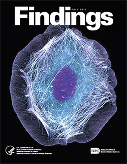
It’s back! Check out the new issue of Findings magazine.

Findings Magazine Cover for Fall 2017.Findings presents cutting-edge research from scientists in diverse biomedical fields. The articles are aimed at high school students with the goal of making science—and the people who do it—interesting and exciting, and to inspire young readers to pursue careers in biomedical research. In addition to putting a face on science, Findings offers activities such as quizzes and crossword puzzles and, in its online version, video interviews with scientists.

The Fall 2017 issue profiles Yale University biologist Enrique De La Cruz, who studies how actin—a protein chain that supports cell structure—breaks so easily. Also profiled is University of California, Berkeley, biologist Rebecca Heald and her study of developmental factors that control an animal’s size.

This issue also features:

  • A virtual reality program designed to help burn patients manage pain
  • The promise of gene therapy for glaucoma
  • The many ways scientists categorize the biological world using “omics”
  • What researchers know—and don’t know—about how general anesthetics work
  • How animation helps researchers visualize interactions between biological molecules
  • How cells use sugary outer coatings to distinguish friend from foe
  • What makes our tissues stiff, squishy, solid, or see-through (hint: its initials are ECM)
  • How super-powerful microscopes are revealing views of biology never possible before

View Findings online, or order a print copy (classroom sets of up to 30 copies are available for educators).

Happy Birthday, BioBeat

0 comments

This month, our blog that highlights NIGMS-funded research turns four years old! For each candle, we thought we’d illuminate an aspect of the blog to offer you, our reader, an insider’s view.
Who are we?

Over the years, the editorial team has included onsite science writers, office interns, staff scientists and guest authors from universities. Kathryn, who’s a regular contributor, writes entirely from her home office. Chris, who has a Ph.D. in neuroscience and now manages the blog, used to do research in a lab. Alisa has worked in NIGMS’ Bethesda-based office the longest: 22 years! She and I remember when we first launched Biomedical Beat as an e-newsletter in 2005. You can read more about each of the writers on the contributors page and if you know someone who’s considering a career in science communications, tell them to drop us a line.

How do we come up with the stories?

We get our story ideas from a range of sources. For instance, newspaper articles about an experimental pest control strategy in Florida and California prompted us to write about NIGMS-funded studies exploring the basic science of the technique. A beautiful visual from a grantee’s institution inspired a short post on tissue regeneration research. And an ongoing conversation with NIGMS scientific staff about the important role of research organisms in biological studies sparked the idea for a playful profile of one such science superstar.

A big change in our storytelling has been shifting the focus from a single finding to broader progress in a lab or field. So instead of reporting on a study just published in a scientific journal, we may write about the scientist’s career path or showcase a collection of recent findings in that particular field. These approaches help us demonstrate that scientific understanding usually progresses through the slow and steady work undertaken by many labs.

What are our favorite posts?

I polled the writers on posts they liked, and the list is really long! Here are the top picks.


Four Ways Inheritance Is More Complex Than Mendel Knew


The Endoplasmic Reticulum: Networking in the Cell


Interview With a Scientist: Janet Iwasa, Molecular Animator


From Basic Research to Bioelectric Medicine


An Insider’s Look at Life: Magnified, an Airport Exhibit of Stunning Microscopy Images

What are your favorite posts?

We regularly review data about the number of times a blog post has been viewed to identify the ones that interest readers the most. That information also helps guide our decisions about other topics to feature on the blog. The Cool Image posts are among the most popular! Below are some other chart-topping posts.


Our Complicated Relationship With Viruses


The Proteasome: The Cells Trash Processor in Action


Demystifying General Anesthetics


Meet Sarkis Mazmanian and the Bacteria That Keep Us Healthy


5 Reasons Biologists Love Math

We always like hearing from readers! If there’s a basic biomedical research topic you’d like us to write about, or if you have feedback on a story or the blog in general, please leave your suggestions in the comment field below.

Chasing Fireflies—and Better Cellular Imaging Techniques

0 comments
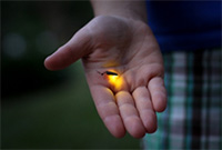
A glowing firefly sitting in a person's open palm. Firefly. Credit: Stock photo.

The yellow-green glow from this summer’s fireflies teased my kids across the yard. Max and Stella zigzagged the grass, occasionally jumping into the air to cup a firefly in their hands and then proudly shouting, “I got one!”

Chasing fireflies on a summer night is a childhood rite of passage for many, including Nathan Shaner who grew up in New Jersey. “It was one of my favorite things about summer,” he recalls. “I’d catch them with my hands—I’d never jar them.”

Today, Shaner studies the science of bioluminescence, which gives fireflies and many other organisms the natural ability to emit light. His goal is to make bright bioluminescent tags that he and other scientists can use to study living cells in greater detail. “There’s this very beautiful thing that evolved in nature, and we can use it to enable new discoveries,” he says.

Thousands of organisms glow as a way to communicate, spook predators, lure prey or attract mates. There are a few terrestrial examples, such as fireflies, glowworm insect larvae and foxfire fungi, and many more aquatic ones, including types of marine plankton, fish, jellyfish, shrimp, squid and sea urchins. One research team estimated nearly three quarters of sea life have bioluminescent capabilities.

Continue reading “Chasing Fireflies—and Better Cellular Imaging Techniques”

Cool Image: Inside a Biofilm Build-up

1 comment
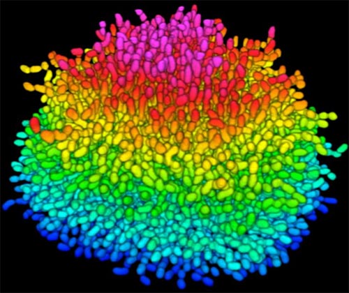
A growing Vibrio cholerae biofilm.

A growing Vibrio cholerae biofilm. Each slightly curved comma shape represents an individual bacterium from assembled confocal microscopy images. Different colors show each bacterium’s position in the biofilm in relation to the surface on which the film is growing. Credit: Jing Yan, Ph.D., and Bonnie Bassler, Ph.D., Department of Molecular Biology, Princeton University, Princeton, NJ.

Bacteria use many methods to overcome threats in their environment. One of these ways is forming colonies called biofilms on surfaces of objects. Often appearing like the bubble-shaped fortress represented in this image, biofilms enable bacteria to withstand attacks, compete for space and survive fluctuations in nutrient supply. Bacteria aggregated within biofilms inside our bodies, for example, resist antibiotic therapy more effectively than free swimming cells, making infections difficult to treat. On the other hand, biofilms are also useful for making microbial fuel cells and for waste-water treatment. Learning how biofilms work, therefore, could provide essential tools in our ongoing battle against disease-causing agents and in our efforts to harness beneficial bacterial behaviors. Researchers are now using new imaging techniques to watch how biofilms grow, cell by cell, and to identify more effective ways of disrupting or fostering them.

Until now, poor imaging resolution meant that scientists could not see what individual bacteria in the films are up to as the biofilms grow. The issue is that bacteria are tiny, making imaging each cell, as well as the ability to distinguish each cell in the biofilm community, problematic. Continue reading “Cool Image: Inside a Biofilm Build-up”

Interview With a Scientist: Thomas O’Halloran, Metal Maestro

2 comments

Inside our bodies is a surprising amount of metal. Not enough to set off the scanners at the airport or make us rich, but enough to fill each of our cells with billions of metal ions, including calcium, iron, copper and zinc. These ions facilitate critical biological functions.

However, too much of any metal can be toxic, while too little can cause disease. Our cells carefully monitor and control their metal content using a whole series of proteins that bind, sense and transport metal ions.

Keeping tabs on why and how metals flow into and out of our cells is a passion of Thomas O’Halloran Exit icon, professor of chemistry and molecular biosciences at Northwestern University in Illinois. For the past three decades, O’Halloran has investigated how fluctuations in the amount of metal ions inside cells influence gene expression, cell growth and other vital functions. Using a variety of approaches, he has uncovered new types of proteins that bind metal ions and investigated the role that imbalances in these ions play in a number of disease-related physiological processes.

One recent focus of his studies has been how zinc regulates oocyte (egg cell) maturation and fertilization. Ultimately, his work could help us better understand infertility, cancer and certain bacterial infections.

There’s an “Ome” for That

1 comment
In the 13 years since the sequencing of the human genome, the list of “omes” has proliferated. Drop us a comment with your favorite ome—we may feature it in a follow-up post next month.

Have you ever collected coins, cards, toy trains, stuffed animals? Did you feel the need to complete the set? If so, then you may be a completist. A completist will go to great lengths to acquire a complete set of something.

Scientists can also be completists who are inspired to identify and catalog every object in a particular field to further our understanding of it. For example, a comprehensive parts list of the human body—and of other organisms that are important in biomedical research—could aid in the development of novel treatments for diseases in the same way that a parts list for a car enables auto mechanics to build or repair a vehicle.

More than 15 years ago, scientists figured out how to catalog every gene in the human body. In the years since, rapid advances in technology and computational tools have allowed researchers to begin to categorize numerous aspects of the biological world. There’s actually a special way to name these collections: Add “ome” to the end of the class of objects being compiled. So, the complete set of genes in the body is called the “genome,” and the complete set of proteins is called the “proteome.”

Below are three -omes that NIH-funded scientists work with to understand human health.

Genome

Illustration of the entire outer shell of the bacteriophage MS2. Credit: Wikimedia Commons, Naranson.

The genome is the original -ome. In 1976, Belgium scientists identified all 3,569 DNA bases—the As, Cs, Gs and Ts that make up DNA’s code—in the genes of bacteriophage MS2, immortalizing this bacteria-infecting virus as possessing the first fully sequenced genome.

Over the next two decades, a small handful of additional genomes from other microorganisms followed. The first animal genome was completed in 1998. Just 5 years later, scientists identified all 3.2 billion DNA bases in the human genome, representing the work of more than 1,000 researchers from six countries over a period of 13 years. Continue reading “There’s an “Ome” for That”

Metals in Medicine

4 comments

An exhibit called “Minerals in Medicine” opened at the NIH Clinical Center last month (see slideshow). The display features a fascinating overview of how dozens of minerals are used to create drugs and medical instruments useful in treating disease and maintaining health. The minerals ranged from commonplace ones like quartz, which is used to make medical instruments, to more exotic ones like hubnerite, a source of the metal tungsten, which is used in radiation shielding.

Inspired by this collection, which is co-sponsored by NIH and the Smithsonian Institution, we highlight here examples of “Metals in Medicine.”

Copper and Fat Metabolism

Fluorescent imaging of copper in white fat cells from mice.

Fluorescent imaging of copper in white fat cells from mice. The left panel shows fat cells with normal levels of copper, and the right panel shows fat cells deficient in copper. Credit: Lakshmi Krishnamoorthy and Joseph Cotruvo Jr., University of California, Berkeley.

What does a metal like copper have to do with our ability to breakdown fat? Researchers explored this question by observing mice with Wilson’s disease—a rare, inherited condition that causes copper to accumulate in the liver, brain and other vital organs. The mice with the condition usually have larger deposits of fat compared to healthy mice. To confirm that fat metabolism is somehow compromised in these mice, the researchers treated them with a drug that induces the breakdown of fat. And indeed they found that less fat was metabolized in mice with the disease.

In an effort to investigate what role copper may be playing in fat metabolism, the researchers examined adipose tissue, or fat, cells under a microscope to track the metal’s interactions with various proteins in the cell. They discovered that copper inhibits an enzyme called PDE3. This enzyme usually prevents another enzyme called cAMP from helping to break down fat. The researchers concluded that copper actually promotes fat metabolism. This work shows that transition metal nutrients can play signaling roles, which has been previously thought to be restricted to alkali and alkaline earth metals like sodium, potassium and calcium.

Continue reading “Metals in Medicine”

Exploring the Evolution of Spider Venom to Improve Human Health

1 comment
Brown recluse

Female brown recluse spider. Credit Matt Bertone, North Carolina State University.

This Halloween, you’re not likely to see many trick-or-treaters dressed as spiders. Google Trends pegs “Spider” as the 87th most searched-for Halloween costume, right between “Hippie” and “The Renaissance.” But don’t let your guard down. Spiders are everywhere.

“I grew up on a farm in Indiana and had the luxury of exploring and turning over rocks and being curious. Any feelings of being grossed out by spiders were rapidly replaced by my feelings of awe for how amazing and diverse these creatures are.”– Greta Binford”

More than 46,000 species of spiders creepy crawl across the globe, on every continent except Antarctica. Each species produces a venom composed of an average of 500 distinct toxins, putting the conservative estimate of unique venom compounds at more than 22 million. This staggering diversity of venoms, collectively referred to as the venome, has only begun to be explored. Continue reading “Exploring the Evolution of Spider Venom to Improve Human Health”